

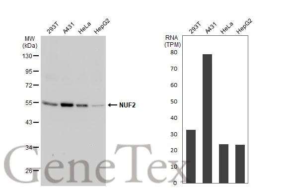
Various whole cell extracts (30 μg) were separated by 10% SDS-PAGE, and the membrane was blotted with NUF2 antibody (GTX110734) diluted at 1:1000. The HRP-conjugated anti-rabbit IgG antibody (GTX213110-01) was used to detect the primary antibody. Corresponding RNA expression data for the same cell lines are based on Human Protein Atlas program.
 were separated by 10% SDS-PAGE, and the membrane was blotted with NUF2 antibody (GTX110734) diluted at 1:1000. The HRP-conjugated anti-rabbit IgG antibody (GTX213110-01) was used to detect the primary antibody.")
![NUF2 antibody detects NUF2 protein at kinetochore by immunofluorescent analysis. Sample: HeLa cells were fixed in 4% paraformaldehyde at RT for 15 min. Green: NUF2 stained by NUF2 antibody (GTX110734) diluted at 1:1000. Red: alpha Tubulin, a cytoskeleton marker, stained by alpha Tubulin antibody [GT114] (GTX628802) diluted at 1:500. Blue: Fluoroshield with DAPI (GTX30920).](https://www.genetex.com/upload/website/prouct_img/normal/GTX110734/GTX110734_43621_20200205_ICC_IF_w_23060500_987.webp "NUF2 antibody detects NUF2 protein at kinetochore by immunofluorescent analysis. Sample: HeLa cells were fixed in 4% paraformaldehyde at RT for 15 min. Green: NUF2 stained by NUF2 antibody (GTX110734) diluted at 1:1000. Red: alpha Tubulin, a cytoskeleton marker, stained by alpha Tubulin antibody [GT114] (GTX628802) diluted at 1:500. Blue: Fluoroshield with DAPI (GTX30920).")
 of paraformaldehyde-fixed U2OS, using NUF2 (GTX110734) antibody (Geen) at 1:500 dilution. Alpha-tubulin filaments were labeled with GTX11304 (Red) at 1:2000.")
Various whole cell extracts (30 μg) were separated by 10% SDS-PAGE, and the membrane was blotted with NUF2 antibody (GTX110734) diluted at 1:1000. The HRP-conjugated anti-rabbit IgG antibody (GTX213110-01) was used to detect the primary antibody. Corresponding RNA expression data for the same cell lines are based on Human Protein Atlas program.
NUF2 antibody
GTX110734
ApplicationsImmunoFluorescence, Western Blot, ImmunoCytoChemistry
Product group Antibodies
ReactivityHuman
TargetNUF2
Overview
- SupplierGeneTex
- Product NameNUF2 antibody
- Delivery Days Customer9
- Application Supplier NoteWB: 1:500-1:3000. ICC/IF: 1:100-1:1000. *Optimal dilutions/concentrations should be determined by the researcher.Not tested in other applications.
- ApplicationsImmunoFluorescence, Western Blot, ImmunoCytoChemistry
- CertificationResearch Use Only
- ClonalityPolyclonal
- Concentration0.52 mg/ml
- ConjugateUnconjugated
- Gene ID83540
- Target nameNUF2
- Target descriptionNUF2 component of NDC80 kinetochore complex
- Target synonymsCDCA1, CT106, NUF2R, kinetochore protein Nuf2, NDC80 kinetochore complex component NUF2, NUF2, NDC80 kinetochore complex component, homolog, cancer/testis antigen 106, cell division cycle associated 1, cell division cycle-associated protein 1, hNuf2, hNuf2R, hsNuf2
- HostRabbit
- IsotypeIgG
- Protein IDQ9BZD4
- Protein NameKinetochore protein Nuf2
- Scientific DescriptionThis gene encodes a protein that is highly similar to yeast Nuf2, a component of a conserved protein complex associated with the centromere. Yeast Nuf2 disappears from the centromere during meiotic prophase when centromeres lose their connection to the spindle pole body, and plays a regulatory role in chromosome segregation. The encoded protein is found to be associated with centromeres of mitotic HeLa cells, which suggests that this protein is a functional homolog of yeast Nuf2. Alternatively spliced transcript variants that encode the same protein have been described. [provided by RefSeq]
- ReactivityHuman
- Storage Instruction-20°C or -80°C,2°C to 8°C
- UNSPSC41116161
Datasheet
Related products


Product group Antibodies
Anti-NUF2 AntibodyHPA059692
ApplicationsImmunoCytoChemistry
ReactivityHuman
TargetNUF2
- SizePrice

Product group Antibodies
Anti-NUF2 Antibody Picoband(r)A03788-2-CARRIER-FREE
ApplicationsWestern Blot, ELISA
ReactivityHuman
TargetNUF2
- SizePrice
Product group Antibodies

CDCA1 / NUF2 AntibodyLS-C676195
ApplicationsWestern Blot, ELISA, ImmunoHistoChemistry, ImmunoHistoChemistry Paraffin
ReactivityHuman
TargetNUF2
- SizePrice
Product group Antibodies
NUF2 AntibodyCSB-PA787491
ApplicationsWestern Blot, ELISA
ReactivityHuman
TargetNUF2
- SizePrice
Product group Antibodies
Nuf2 Polyclonal AntibodyCAC10816
ApplicationsWestern Blot, ELISA, ImmunoHistoChemistry
TargetNUF2
- SizePrice
Product group Antibodies
References
NUF2 Polyclonal AntibodyBS-7714R
ApplicationsFlow Cytometry, ImmunoFluorescence, ELISA, ImmunoCytoChemistry, ImmunoHistoChemistry, ImmunoHistoChemistry Frozen, ImmunoHistoChemistry Paraffin
ReactivityBovine, Canine, Equine, Human, Mouse, Porcine, Rabbit, Rat
TargetNUF2
- SizePrice
![Immunoprecipitation of NUF2 protein from HeLa whole cell extracts using 5 μg of NUF2 antibody [GT644] (GTX632184) . Western blot analysis was performed using NUF2 antibody [GT644] (GTX632184) diluted at 1:500. EasyBlot anti-Mouse IgG (GTX221667-01) was used as a secondary reagent.](https://www.genetex.com/upload/website/prouct_img/normal/GTX632184/GTX632184_42044_20160715_IP_w_23061202_330.webp)
Product group Antibodies
NUF2 antibody [GT644]GTX632184
ApplicationsImmunoPrecipitation, Western Blot
ReactivityHuman
TargetNUF2
- SizePrice
![NUF2 antibody [GT312] detects NUF2 protein at nucleus by immunohistochemical analysis. Sample: Paraffin-embedded human breast carcinoma. NUF2 stained by NUF2 antibody [GT312] (GTX632247) diluted at 1:500. Antigen Retrieval: Citrate buffer, pH 6.0, 15 min](https://www.genetex.com/upload/website/prouct_img/normal/GTX632247/GTX632247_44706_20230303_IHC-P_23032819_979.webp)
Product group Antibodies
NUF2 antibody [GT312]GTX632247
ApplicationsImmunoPrecipitation, Western Blot, ImmunoHistoChemistry, ImmunoHistoChemistry Paraffin
ReactivityHuman
TargetNUF2
- SizePrice