

![ODC-1 (Ornithine Decarboxylase-1)(ODC1/487), Biotin conjugate, 0.1mg/mL [26628-22-8]](https://biotium.com/wp-content/uploads/2016/12/BNUB0487-1-1.jpg "ODC-1 (Ornithine Decarboxylase-1)(ODC1/487), Biotin conjugate, 0.1mg/mL [26628-22-8]")
ODC-1 (Ornithine Decarboxylase-1)(ODC1/487), Biotin conjugate, 0.1mg/mL [26628-22-8]
BNCB0487
ApplicationsFlow Cytometry, ImmunoFluorescence
Product group Antibodies
ReactivityBovine, Human, Mouse, Rat
TargetODC1
Overview
- SupplierBiotium
- Product NameODC-1 (Ornithine Decarboxylase-1)(ODC1/487), Biotin conjugate, 0.1mg/mL [26628-22-8]
- Delivery Days Customer9
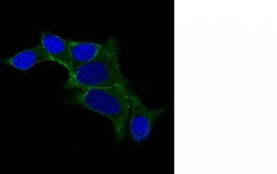
- ApplicationsFlow Cytometry, ImmunoFluorescence
- CAS Number26628-22-8
- CertificationResearch Use Only
- ClonalityMonoclonal
- Clone IDODC1/487
- Concentration0.1 mg/ml
- ConjugateBiotin
- Gene ID4953
- Target nameODC1
- Target descriptionornithine decarboxylase 1
- Target synonymsBABS, NEDBA, NEDBIA, ODC, ornithine decarboxylase
- HostMouse
- IsotypeIgG2a
- Protein IDP11926
- Protein NameOrnithine decarboxylase
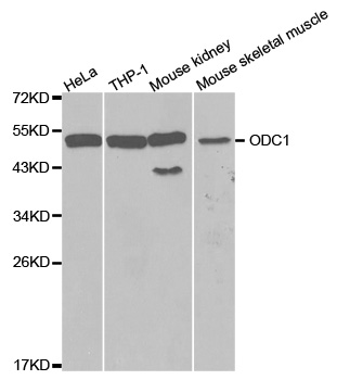
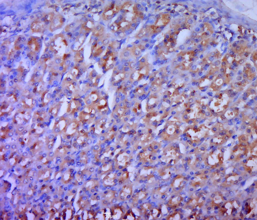
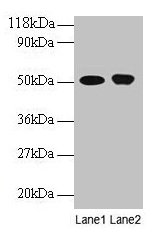
- Scientific DescriptionRecognizes a 53 kDa protein, identified as the Ornithine Decarboxylase (ODC-1). ODC is the initial and rate-limiting enzyme in the biosynthetic pathway of polyamines and is involved in the conversion of ornithine to putrescine. The biological activity of ODC-1 is rapidly induced in response to virtually all agents known to promote cell proliferation including hormones, drugs, growth factors, mitogens, and tumor promoters. Reportedly, ODC mRNA levels are elevated in lung carcinomas as well as in colon adenomas and carcinomas. ODC activity in colorectal carcinomas is greater than those in adenomas and normal mucosa.Primary antibodies are available purified, or with a selection of fluorescent CF® Dyes and other labels. CF® Dyes offer exceptional brightness and photostability. Note: Conjugates of blue fluorescent dyes like CF®405S and CF®405M are not recommended for detecting low abundance targets, because blue dyes have lower fluorescence and can give higher non-specific background than other dye colors.
- SourceAnimal
- ReactivityBovine, Human, Mouse, Rat
- Storage Instruction2°C to 8°C,RT
- UNSPSC41116161
MSDS
Related products

Product group Antibodies
Anti-ODC1 Antibody Picoband(r)A03138-1-CARRIER-FREE
ApplicationsWestern Blot, ELISA
ReactivityHuman, Mouse, Rat
TargetODC1
- SizePrice
Product group Antibodies
Anti-ODC1 Antibody144-01948
ApplicationsWestern Blot, ImmunoHistoChemistry
ReactivityHuman, Mouse, Rat
TargetODC1
- SizePrice
Product group Antibodies

Anti-ODC1 [ODC1-2]AB03819-10.0
ApplicationsWestern Blot, ELISA, ImmunoHistoChemistry
ReactivityHuman
TargetODC1
- SizePrice
Product group Antibodies
Anti-ODC1 AntibodyA30530
ApplicationsWestern Blot, ImmunoHistoChemistry
ReactivityHuman, Mouse, Rat
- SizePrice
Product group Antibodies
References
ODC1 Polyclonal AntibodyBS-1294R
ApplicationsImmunoFluorescence, Western Blot, ELISA, ImmunoCytoChemistry, ImmunoHistoChemistry, ImmunoHistoChemistry Frozen, ImmunoHistoChemistry Paraffin
ReactivityBovine, Canine, Equine, Human, Mouse, Porcine, Rat
TargetODC1
- SizePrice
Product group Antibodies
Odc1 Polyclonal AntibodyCAC07342
ApplicationsImmunoFluorescence, Western Blot, ELISA, ImmunoHistoChemistry
TargetODC1
- SizePrice
Product group Antibodies
ODC1 AntibodyCSB-PA00535A0RB
ApplicationsImmunoFluorescence, Western Blot, ELISA, ImmunoHistoChemistry
ReactivityHuman
TargetODC1
- SizePrice
![Various whole cell extracts (30 μg) were separated by 10% SDS-PAGE, and the membrane was blotted with ODC antibody [N3C3] (GTX101521) diluted at 1:1000. The HRP-conjugated anti-rabbit IgG antibody (GTX213110-01) was used to detect the primary antibody, and the signal was developed with Trident ECL plus-Enhanced. Corresponding RNA expression data for the same cell lines are based on Human Protein Atlas program.](https://www.genetex.com/upload/website/prouct_img/normal/GTX101521/GTX101521_44804_20221202_WB_TPM_watermark_22122722_565.webp)
Product group Antibodies
ODC antibody [N3C3]GTX101521
ApplicationsWestern Blot, ImmunoHistoChemistry, ImmunoHistoChemistry Paraffin
ReactivityHuman, Mouse, Rat
TargetODC1
- SizePrice
Product group Antibodies

ODC1 / Ornithine Decarboxylase AntibodyLS-C331782
ApplicationsWestern Blot, ImmunoHistoChemistry
ReactivityHuman, Mouse
TargetODC1
- SizePrice