

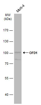
Whole cell extract (30 μg) was separated by 7.5% SDS-PAGE, and the membrane was blotted with OFD1 antibody (GTX110010) diluted at 1:5000. The HRP-conjugated anti-rabbit IgG antibody (GTX213110-01) was used to detect the primary antibody.
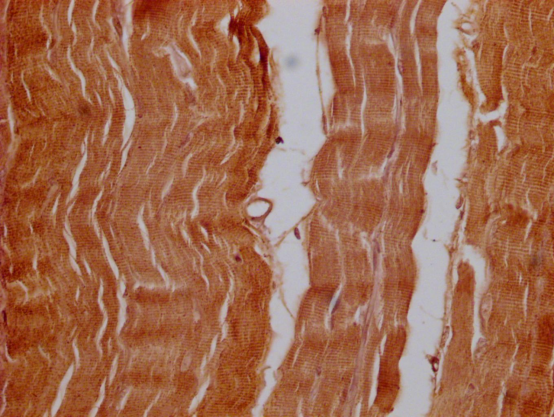
 dilution: 1:500.
Antigen Retrieval: Trilogy? (EDTA based, pH 8.0) buffer, 15min")
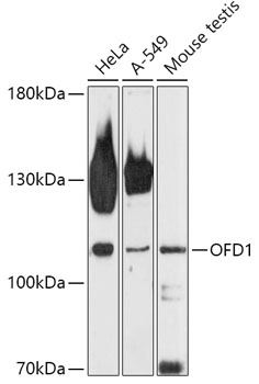
 were separated by 7.5% SDS-PAGE, and the membrane was blotted with OFD1 antibody (GTX110010) diluted at 1:1000. The HRP-conjugated anti-rabbit IgG antibody (GTX213110-01) was used to detect the primary antibody.")


Whole cell extract (30 μg) was separated by 7.5% SDS-PAGE, and the membrane was blotted with OFD1 antibody (GTX110010) diluted at 1:5000. The HRP-conjugated anti-rabbit IgG antibody (GTX213110-01) was used to detect the primary antibody.
OFD1 antibody
GTX110010
ApplicationsWestern Blot, ImmunoHistoChemistry, ImmunoHistoChemistry Paraffin
Product group Antibodies
ReactivityHuman, Mouse
TargetOFD1
Overview
- SupplierGeneTex
- Product NameOFD1 antibody
- Delivery Days Customer9
- Application Supplier NoteWB: 1:1000-1:10000. IHC-P: 1:100-1:1000. *Optimal dilutions/concentrations should be determined by the researcher.Not tested in other applications.
- ApplicationsWestern Blot, ImmunoHistoChemistry, ImmunoHistoChemistry Paraffin
- CertificationResearch Use Only
- ClonalityPolyclonal
- Concentration1.21 mg/ml
- ConjugateUnconjugated
- Gene ID8481
- Target nameOFD1
- Target descriptionOFD1 centriole and centriolar satellite protein
- Target synonyms71-7A, CXorf5, JBTS10, RP23, SGBS2, centriole and centriolar satellite protein OFD1, Joubert syndrome type 10, oral-facial-digital syndrome 1 protein, protein 71-7A
- HostRabbit
- IsotypeIgG
- Protein IDO75665
- Protein NameCentriole and centriolar satellite protein OFD1
- Scientific DescriptionThis gene is located on the X chromosome and encodes a centrosomal protein. A knockout mouse model has been used to study the effect of mutations in this gene. The mouse gene is also located on the X chromosome, however, unlike the human gene it is not subject to X inactivation. Mutations in this gene are associated with oral-facial-digital syndrome type I and Simpson-Golabi-Behmel syndrome type 2. Many pseudogenes have been identified; a single pseudogene is found on chromosome 5 while as many as fifteen have been found on the Y chromosome. Alternatively spliced transcripts have been described for this gene but the biological validity of these transcripts has not been determined. [provided by RefSeq]
- ReactivityHuman, Mouse
- Storage Instruction-20°C or -80°C,2°C to 8°C
- UNSPSC41116161
Datasheet
Related products
Product group Antibodies
Anti-OFD1 AntibodyA308250
ApplicationsWestern Blot
ReactivityHuman, Mouse
- SizePrice

Product group Antibodies
Anti-OFD1 Antibody Picoband(r)A02955-1-CARRIER-FREE
ApplicationsFlow Cytometry, ImmunoFluorescence, Western Blot, ImmunoCytoChemistry
ReactivityHuman, Mouse, Rat
TargetOFD1
- SizePrice
Product group Antibodies
Anti-OFD1 Antibody107-10889
ApplicationsWestern Blot, ImmunoHistoChemistry, ImmunoHistoChemistry Paraffin
ReactivityHuman
TargetOFD1
- SizePrice
Product group Antibodies
OFD1 Polyclonal AntibodyCAC13142
ApplicationsImmunoFluorescence, Western Blot, ELISA
ReactivityMouse, Rat
TargetOFD1
- SizePrice
Product group Antibodies
OFD1 AntibodyCSB-PA016289LA01HU
ApplicationsImmunoFluorescence, Western Blot, ELISA, ImmunoHistoChemistry
ReactivityHuman
TargetOFD1
- SizePrice
Product group Antibodies

OFD1 AntibodyLS-C499455
ApplicationsImmunoFluorescence, Western Blot, ELISA, ImmunoHistoChemistry
ReactivityHuman, Mouse
TargetOFD1
- SizePrice

Product group Antibodies
Anti-OFD1 AntibodyHPA031103
ApplicationsWestern Blot
ReactivityHuman
TargetOFD1
- SizePrice