

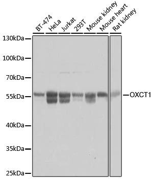
WB analysis of various sample lysates using GTX32770 OXCT1 antibody. Dilution : 1:3000 Loading : 25μg per lane
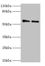
 and knockout (KO) HeLa cell lysate using GTX32770 OXCT1 antibody. Dilution : 1:3000 Loading : 25μg per lane")


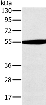
WB analysis of various sample lysates using GTX32770 OXCT1 antibody. Dilution : 1:3000 Loading : 25μg per lane
OXCT1 antibody
GTX32770
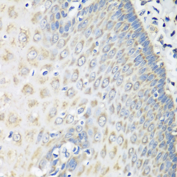
ApplicationsWestern Blot, ImmunoHistoChemistry, ImmunoHistoChemistry Paraffin
Product group Antibodies
ReactivityHuman, Mouse, Rat
TargetOXCT1
Overview
- SupplierGeneTex
- Product NameOXCT1 antibody
- Delivery Days Customer9
- Application Supplier NoteWB: 1:500 - 1:2000. IHC-P: 1:50 - 1:100. *Optimal dilutions/concentrations should be determined by the researcher.Not tested in other applications.
- ApplicationsWestern Blot, ImmunoHistoChemistry, ImmunoHistoChemistry Paraffin
- CertificationResearch Use Only
- ClonalityPolyclonal
- ConjugateUnconjugated
- Gene ID5019
- Target nameOXCT1
- Target description3-oxoacid CoA-transferase 1
- Target synonymsOXCT, SCOT, succinyl-CoA:3-ketoacid coenzyme A transferase 1, mitochondrial, 3-oxoacid CoA transferase, SCOT-s, epididymis secretory sperm binding protein, somatic-type succinyl-CoA:3-oxoacid CoA-transferase, succinyl CoA:3-oxoacid CoA transferase, succinyl-CoA:3-ketoacid-CoA transferase
- HostRabbit
- IsotypeIgG
- Protein IDP55809
- Protein NameSuccinyl-CoA:3-ketoacid coenzyme A transferase 1, mitochondrial
- Scientific DescriptionThis gene encodes a member of the 3-oxoacid CoA-transferase gene family. The encoded protein is a homodimeric mitochondrial matrix enzyme that plays a central role in extrahepatic ketone body catabolism by catalyzing the reversible transfer of coenzyme A from succinyl-CoA to acetoacetate. Mutations in this gene are associated with succinyl CoA:3-oxoacid CoA transferase deficiency. [provided by RefSeq, Jul 2008]
- ReactivityHuman, Mouse, Rat
- Storage Instruction-20°C or -80°C,2°C to 8°C
- UNSPSC41116161
Datasheet
Related products
Product group Antibodies
OXCT1 AntibodyCSB-PA017306ESR1HU
ApplicationsWestern Blot, ELISA
ReactivityHuman, Mouse
TargetOXCT1
- SizePrice

Product group Antibodies
Anti-OXCT1/SCOT Antibody Picoband(r)A07229-1-CARRIER-FREE
ApplicationsFlow Cytometry, ImmunoFluorescence, Western Blot, ELISA, ImmunoCytoChemistry
ReactivityHuman, Monkey, Mouse, Rat
TargetOXCT1
- SizePrice

Product group Antibodies
Anti-OXCT1 AntibodyHPA012047
ApplicationsWestern Blot, ImmunoCytoChemistry, ImmunoHistoChemistry
ReactivityHuman, Mouse, Rat
TargetOXCT1
- SizePrice
Product group Antibodies

OXCT1 AntibodyLS-C409675
ApplicationsWestern Blot, ImmunoHistoChemistry
ReactivityHuman, Mouse, Rat
TargetOXCT1
- SizePrice
Product group Antibodies
OXCT1 Polyclonal AntibodyCAC13855
ApplicationsWestern Blot, ELISA, ImmunoHistoChemistry
TargetOXCT1
- SizePrice
Product group Antibodies
Anti-OXCT1 Antibody144-08139
ApplicationsWestern Blot, ImmunoHistoChemistry
ReactivityHuman, Mouse, Rat
TargetOXCT1
- SizePrice
Product group Antibodies
OXCT1 Polyclonal AntibodyBS-55159R
ApplicationsWestern Blot, ImmunoHistoChemistry, ImmunoHistoChemistry Paraffin
ReactivityHuman, Mouse, Rat
TargetOXCT1
- SizePrice