

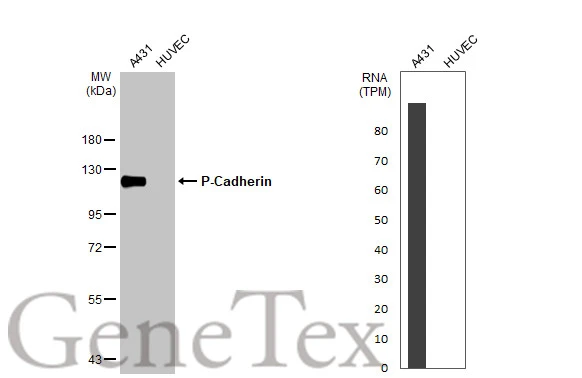
Various whole cell extracts (30 μg) were separated by 7.5% SDS-PAGE, and the membrane was blotted with P-Cadherin antibody (GTX113648) diluted at 1:5000. The HRP-conjugated anti-rabbit IgG antibody (GTX213110-01) was used to detect the primary antibody.Corresponding RNA expression data for the same cell lines are based on Human Protein Atlas program.
 antibody at 1:100 dilution.
Antigen Retrieval: Trilogy? (EDTA based, pH 8.0) buffer, 15min")
Various whole cell extracts (30 μg) were separated by 7.5% SDS-PAGE, and the membrane was blotted with P-Cadherin antibody (GTX113648) diluted at 1:5000. The HRP-conjugated anti-rabbit IgG antibody (GTX213110-01) was used to detect the primary antibody.Corresponding RNA expression data for the same cell lines are based on Human Protein Atlas program.
P-Cadherin antibody
GTX113648
ApplicationsWestern Blot, ImmunoHistoChemistry, ImmunoHistoChemistry Paraffin
Product group Antibodies
ReactivityHuman, Mouse
TargetCDH3
Overview
- SupplierGeneTex
- Product NameP-Cadherin antibody
- Delivery Days Customer9
- Application Supplier NoteWB: 1:1000-1:10000. IHC-P: 1:100-1:1000. *Optimal dilutions/concentrations should be determined by the researcher.Not tested in other applications.
- ApplicationsWestern Blot, ImmunoHistoChemistry, ImmunoHistoChemistry Paraffin
- CertificationResearch Use Only
- ClonalityPolyclonal
- Concentration1 mg/ml
- ConjugateUnconjugated
- Gene ID1001
- Target nameCDH3
- Target descriptioncadherin 3
- Target synonymsCDHP, HJMD, PCAD, cadherin-3, cadherin 3, type 1, P-cadherin (placental), calcium-dependent adhesion protein, placental
- HostRabbit
- IsotypeIgG
- Protein IDP22223
- Protein NameCadherin-3
- Scientific DescriptionThis gene is a classical cadherin from the cadherin superfamily. The encoded protein is a calcium-dependent cell-cell adhesion glycoprotein comprised of five extracellular cadherin repeats, a transmembrane region and a highly conserved cytoplasmic tail. This gene is located in a six-cadherin cluster in a region on the long arm of chromosome 16 that is involved in loss of heterozygosity events in breast and prostate cancer. In addition, aberrant expression of this protein is observed in cervical adenocarcinomas. Mutations in this gene have been associated with congential hypotrichosis with juvenile macular dystrophy. [provided by RefSeq]
- ReactivityHuman, Mouse
- Storage Instruction-20°C or -80°C,2°C to 8°C
- UNSPSC41116161
Datasheet
Related products
Product group Antibodies
Anti-CDH3 AntibodyA99919
ApplicationsWestern Blot, ELISA, ImmunoHistoChemistry
ReactivityHuman
- SizePrice
Product group Antibodies

Anti-P-Cadherin [6A9]AB02535-1.1
ApplicationsImmunoFluorescence, ImmunoPrecipitation, Western Blot, ImmunoHistoChemistry
ReactivityHuman
TargetCDH3
- SizePrice

Product group Antibodies
Anti-P-Cadherin-3 CDH3-Antibody Picoband(r)A03353-1-CARRIER-FREE
ApplicationsFlow Cytometry, ImmunoFluorescence, Western Blot, ELISA, ImmunoCytoChemistry
ReactivityHuman
TargetCDH3
- SizePrice
Product group Antibodies
Anti-CDH3 Antibody144-63143
ApplicationsWestern Blot, ImmunoHistoChemistry
ReactivityHuman, Mouse, Rat
TargetCDH3
- SizePrice
Product group Antibodies

CDH3 / P-Cadherin AntibodyLS-C749240
ApplicationsWestern Blot, ImmunoHistoChemistry
ReactivityHuman, Mouse, Rat
TargetCDH3
- SizePrice
Product group Antibodies
P-Cadherin (N-terminal region) AntibodyBSM-70369M
ApplicationsWestern Blot
ReactivityHuman, Mouse, Rat
TargetCDH3
- SizePrice
Product group Antibodies
CDH3 AntibodyCSB-PA006358
ApplicationsWestern Blot, ELISA, ImmunoHistoChemistry
ReactivityHuman
TargetCDH3
- SizePrice
Product group Antibodies
ApplicationsWestern Blot, ELISA, ImmunoCytoChemistry, ImmunoHistoChemistry, ImmunoHistoChemistry Frozen, ImmunoHistoChemistry Paraffin
ReactivityMouse
TargetCDH3
- SizePrice
![ICC/IF analysis of BEAS-2B Cells using GTX19350 P-Cadherin antibody [6A9]. Cells were probed without (right) or with(left) an antibody. Green : Primary antibody Blue : Nuclei Red : Actin Fixation : formaldehyde Dilution : 1:20 overnight at 4oC](https://www.genetex.com/upload/website/prouct_img/normal/GTX19350/GTX19350_366_ICC-IF_w_23060620_354.webp)
Product group Antibodies
P-Cadherin antibody [6A9]GTX19350
ApplicationsImmunoFluorescence, ImmunoPrecipitation, Western Blot, ImmunoCytoChemistry, Neutralisation/Blocking
ReactivityHuman, Mouse
TargetCDH3
- SizePrice

Product group Antibodies
Anti-CDH3 AntibodyHPA001767
ApplicationsWestern Blot, ImmunoHistoChemistry
ReactivityHuman
TargetCDH3
- SizePrice