

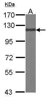
Sample (30 ug of whole cell lysate) A:NIH-3T3 7.5% SDS PAGE GTX100605 diluted at 1:1000
 A: H1299 7.5% SDS PAGE GTX100605 diluted at 1:1000")
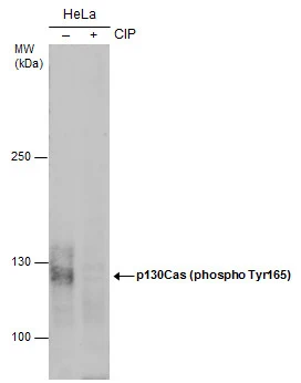
![Non-transfected (–) and transfected (+) 293T whole cell extracts (30 μg) were separated by 7.5% SDS-PAGE, and the membrane was blotted with p130Cas antibody [N2C2], Internal (GTX100605) diluted at 1:500. The HRP-conjugated anti-rabbit IgG antibody (GTX213110-01) was used to detect the primary antibody.](https://www.genetex.com/upload/website/prouct_img/normal/GTX100605/GTX100605_39694_20170518_WB_shRNA_watermark_w_23060100_127.webp "Non-transfected (–) and transfected (+) 293T whole cell extracts (30 μg) were separated by 7.5% SDS-PAGE, and the membrane was blotted with p130Cas antibody [N2C2], Internal (GTX100605) diluted at 1:500. The HRP-conjugated anti-rabbit IgG antibody (GTX213110-01) was used to detect the primary antibody.")
![BCAR1 antibody [N2C2], Internal detects BCAR1 protein at cytoplasm by immunofluorescent analysis. Sample: HeLa cells were fixed in ice-cold MeOH for 5 min. Green: BCAR1 protein stained by BCAR1 antibody [N2C2], Internal (GTX100605) diluted at 1:500. Blue: Hoechst 33342 staining.](https://www.genetex.com/upload/website/prouct_img/normal/GTX100605/GTX100605_39694_IFA_w_23060100_518.webp "BCAR1 antibody [N2C2], Internal detects BCAR1 protein at cytoplasm by immunofluorescent analysis. Sample: HeLa cells were fixed in ice-cold MeOH for 5 min. Green: BCAR1 protein stained by BCAR1 antibody [N2C2], Internal (GTX100605) diluted at 1:500. Blue: Hoechst 33342 staining.")
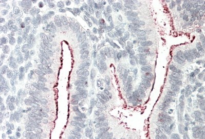
![BCAR1 antibody [N2C2], Internal detects BCAR1 protein at cytoplasm on human breast carcinoma by immunohistochemical analysis. Sample: Paraffin-embedded human breast carcinoma. BCAR1 antibody [N2C2], Internal (GTX100605) diluted at 1:500.
Antigen Retrieval: Trilogy? (EDTA based, pH 8.0) buffer, 15min](https://www.genetex.com/upload/website/prouct_img/normal/GTX100605/GTX100605_39694_20141219_IHC_w_23060100_422.webp "BCAR1 antibody [N2C2], Internal detects BCAR1 protein at cytoplasm on human breast carcinoma by immunohistochemical analysis. Sample: Paraffin-embedded human breast carcinoma. BCAR1 antibody [N2C2], Internal (GTX100605) diluted at 1:500.
Antigen Retrieval: Trilogy? (EDTA based, pH 8.0) buffer, 15min")
![p130Cas antibody [N2C2], Internal detects p130Cas protein expression by immunohistochemical analysis. Sample: Frozen-sectioned adult mouse cerebellum. Green: p130Cas protein stained by p130Cas antibody [N2C2], Internal (GTX100605) diluted at 1:250. Red: NF-H, stained by NF-H antibody [GT114] (GTX634289) diluted at 1:500. Blue: Fluoroshield with DAPI (GTX30920).
Antigen Retrieval: Citrate buffer, pH 6.0, 10 min](https://www.genetex.com/upload/website/prouct_img/normal/GTX100605/GTX100605_41808_20170831_IHC-Fr_M_w_23060100_534.webp "p130Cas antibody [N2C2], Internal detects p130Cas protein expression by immunohistochemical analysis. Sample: Frozen-sectioned adult mouse cerebellum. Green: p130Cas protein stained by p130Cas antibody [N2C2], Internal (GTX100605) diluted at 1:250. Red: NF-H, stained by NF-H antibody [GT114] (GTX634289) diluted at 1:500. Blue: Fluoroshield with DAPI (GTX30920).
Antigen Retrieval: Citrate buffer, pH 6.0, 10 min")
![p130Cas antibody [N2C2], Internal detects p130Cas protein at cytoplasm in mouse brain by immunohistochemical analysis. Sample: Paraffin-embedded mouse brain. p130Cas antibody [N2C2], Internal (GTX100605) diluted at 1:500.
Antigen Retrieval: Citrate buffer, pH 6.0, 15 min](https://www.genetex.com/upload/website/prouct_img/normal/GTX100605/GTX100605_41808_20171024_IHC-P_M_w_23060100_488.webp "p130Cas antibody [N2C2], Internal detects p130Cas protein at cytoplasm in mouse brain by immunohistochemical analysis. Sample: Paraffin-embedded mouse brain. p130Cas antibody [N2C2], Internal (GTX100605) diluted at 1:500.
Antigen Retrieval: Citrate buffer, pH 6.0, 15 min")
![p130Cas antibody [N2C2], Internal detects p130Cas protein at cytoplasm in rat brain by immunohistochemical analysis. Sample: Paraffin-embedded rat brain. p130Cas antibody [N2C2], Internal (GTX100605) diluted at 1:500.
Antigen Retrieval: Citrate buffer, pH 6.0, 15 min](https://www.genetex.com/upload/website/prouct_img/normal/GTX100605/GTX100605_41808_20171024_IHC-P_R_w_23060100_588.webp "p130Cas antibody [N2C2], Internal detects p130Cas protein at cytoplasm in rat brain by immunohistochemical analysis. Sample: Paraffin-embedded rat brain. p130Cas antibody [N2C2], Internal (GTX100605) diluted at 1:500.
Antigen Retrieval: Citrate buffer, pH 6.0, 15 min")
Sample (30 ug of whole cell lysate) A:NIH-3T3 7.5% SDS PAGE GTX100605 diluted at 1:1000
p130Cas antibody [N2C2], Internal
GTX100605
ApplicationsImmunoFluorescence, Western Blot, ImmunoCytoChemistry, ImmunoHistoChemistry, ImmunoHistoChemistry Frozen, ImmunoHistoChemistry Paraffin
Product group Antibodies
ReactivityHuman, Mouse, Rat
TargetBCAR1
Overview
- SupplierGeneTex
- Product Namep130Cas antibody [N2C2], Internal
- Delivery Days Customer9
- Application Supplier NoteWB: 1:500-1:3000. ICC/IF: 1:100-1:1000. IHC-P: 1:100-1:1000. IHC-Fr: 1:100-1:1000. *Optimal dilutions/concentrations should be determined by the researcher.Not tested in other applications.
- ApplicationsImmunoFluorescence, Western Blot, ImmunoCytoChemistry, ImmunoHistoChemistry, ImmunoHistoChemistry Frozen, ImmunoHistoChemistry Paraffin
- CertificationResearch Use Only
- ClonalityPolyclonal
- Concentration0.38 mg/ml
- ConjugateUnconjugated
- Gene ID9564
- Target nameBCAR1
- Target descriptionBCAR1 scaffold protein, Cas family member
- Target synonymsCAS, CAS1, CASS1, CRKAS, P130Cas, breast cancer anti-estrogen resistance protein 1, BCAR1, Cas family scaffold protein, BCAR1, Cas family scaffolding protein, Cas scaffolding protein family member 1, Crk-associated substrate p130Cas
- HostRabbit
- IsotypeIgG
- Protein IDP56945
- Protein NameBreast cancer anti-estrogen resistance protein 1
- Scientific DescriptionBCAR1, or CAS, is an Src (MIM 190090) family kinase substrate involved in various cellular events, including migration, survival, transformation, and invasion (Sawada et al., 2006 [PubMed 17129785]).[supplied by OMIM]
- ReactivityHuman, Mouse, Rat
- Storage Instruction-20°C or -80°C,2°C to 8°C
- UNSPSC41116161
Datasheet
Related products
Product group Antibodies
Anti-BCAR1 AntibodyA82532
ApplicationsWestern Blot, ELISA, ImmunoHistoChemistry
ReactivityHuman
- SizePrice
Product group Antibodies
Anti-Phospho-BCAR1-Y410 Antibody144-50775
ApplicationsWestern Blot
ReactivityHuman, Mouse
TargetBCAR1
- SizePrice
Product group Antibodies

ApplicationsWestern Blot
ReactivityHuman, Mouse
TargetBCAR1
- SizePrice
Product group Antibodies
ApplicationsFlow Cytometry, Western Blot
ReactivityHuman
TargetBCAR1
- SizePrice
Product group Antibodies
BCAR1 AntibodyCSB-PA002593LA01HU
ApplicationsImmunoFluorescence, Western Blot, ELISA, ImmunoHistoChemistry
ReactivityHuman
TargetBCAR1
- SizePrice
Product group Antibodies

Goat anti-BCAR1EB12156
ApplicationsWestern Blot, ELISA, ImmunoHistoChemistry
ReactivityCanine, Human, Mouse, Rat
TargetBCAR1
- SizePrice
Product group Antibodies
BCAR1 Polyclonal AntibodyCAC14981
ApplicationsImmunoFluorescence, Western Blot, ELISA, ImmunoHistoChemistry
TargetBCAR1
- SizePrice

Product group Antibodies
ApplicationsWestern Blot
ReactivityHuman, Mouse, Rat
TargetBCAR1
- SizePrice
Product group Antibodies
p130Cas (phospho Tyr165) antibodyGTX132160
ApplicationsImmunoFluorescence, Western Blot, ImmunoCytoChemistry
ReactivityHuman, Mouse, Rat
TargetBCAR1
- SizePrice
Product group Antibodies
p130Cas (phospho Tyr410) antibodyGTX132162
ApplicationsImmunoFluorescence, Western Blot, ImmunoCytoChemistry
ReactivityHuman
TargetBCAR1
- SizePrice