

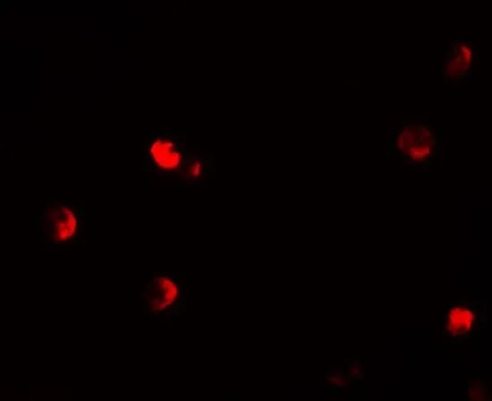
ICC/IF analysis of 293 cells using GTX31859 p57 Kip2 antibody. Working concentration : 20 μg/ml
 1 and (B) 2 μg/ml")
ICC/IF analysis of 293 cells using GTX31859 p57 Kip2 antibody. Working concentration : 20 μg/ml
p57 Kip2 antibody
GTX31859
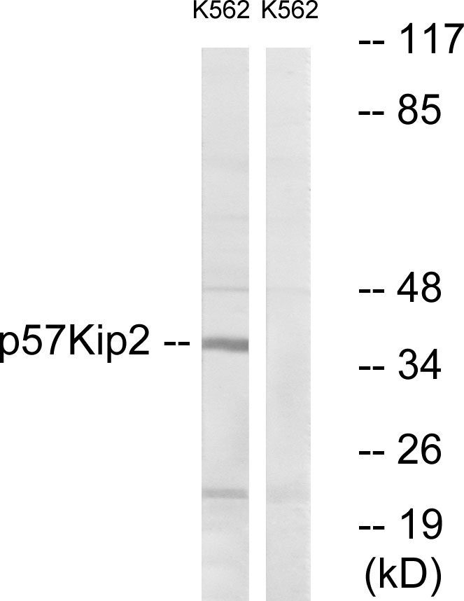
ApplicationsImmunoFluorescence, Western Blot, ELISA, ImmunoCytoChemistry
Product group Antibodies
ReactivityHuman
TargetCDKN1C
Overview
- SupplierGeneTex
- Product Namep57 Kip2 antibody
- Delivery Days Customer9
- Application Supplier NoteWB: 1 - 2 microg/mL. ICC/IF: 20 microg/mL. *Optimal dilutions/concentrations should be determined by the researcher.Not tested in other applications.
- ApplicationsImmunoFluorescence, Western Blot, ELISA, ImmunoCytoChemistry
- CertificationResearch Use Only
- ClonalityPolyclonal
- Concentration1 mg/ml
- ConjugateUnconjugated
- Gene ID1028
- Target nameCDKN1C
- Target descriptioncyclin dependent kinase inhibitor 1C
- Target synonymsBWCR, BWS, KIP2, WBS, p57, p57Kip2, cyclin-dependent kinase inhibitor 1C, cyclin-dependent kinase inhibitor 1C (p57, Kip2), cyclin-dependent kinase inhibitor p57
- HostRabbit
- IsotypeIgG
- Protein IDP49918
- Protein NameCyclin-dependent kinase inhibitor 1C
- Scientific DescriptionThis gene is imprinted, with preferential expression of the maternal allele. The encoded protein is a tight-binding, strong inhibitor of several G1 cyclin/Cdk complexes and a negative regulator of cell proliferation. Mutations in this gene are implicated in sporadic cancers and Beckwith-Wiedemann syndorome, suggesting that this gene is a tumor suppressor candidate. Three transcript variants encoding two different isoforms have been found for this gene. [provided by RefSeq, Oct 2010]
- ReactivityHuman
- Storage Instruction-20°C or -80°C,2°C to 8°C
- UNSPSC41116161
References
- Effect of Butyrate on Collagen Expression, Cell Viability, Cell Cycle Progression and Related Proteins Expression of MG-63 Osteoblastic Cells. Chang MC et al., 2016, PLoS OneRead this paper
Datasheet
Related products
Product group Antibodies
Anti-p57 Kip2 AntibodyA96518
ApplicationsImmunoFluorescence, Western Blot, ELISA
ReactivityHuman, Mouse
- SizePrice


Product group Antibodies
Anti-p57 Kip2/CDKN1C Antibody Picoband(r)A01244-1-CARRIER-FREE
ApplicationsWestern Blot
ReactivityHuman
TargetCDKN1C
- SizePrice
Product group Antibodies
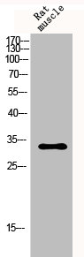
Anti-Mouse/Rat CDKN1C Antibody144-02060
ApplicationsWestern Blot
ReactivityHuman, Mouse, Rat
TargetCDKN1C
- SizePrice
Product group Antibodies

CDKN1C / p57 Kip2 AntibodyLS-C761127
ApplicationsImmunoFluorescence, Western Blot, ImmunoCytoChemistry, ImmunoHistoChemistry
ReactivityHuman, Mouse
TargetCDKN1C
- SizePrice
Product group Antibodies
p57 Kip2/Cdkn1c Recombinant AntibodyBSM-61145R
ApplicationsImmunoFluorescence, ImmunoPrecipitation, Western Blot, ImmunoCytoChemistry, ImmunoHistoChemistry, ImmunoHistoChemistry Frozen, ImmunoHistoChemistry Paraffin
TargetCDKN1C
- SizePrice
Product group Antibodies
CDKN1C AntibodyCSB-PA003684
ApplicationsWestern Blot, ELISA, ImmunoHistoChemistry
ReactivityHuman
TargetCDKN1C
- SizePrice
![IHC-P analysis of human placenta tissue using GTX01862 p57 Kip2 antibody [25B2]. Note nuclear staining for cytotrophoblast and stromal cells of the villi.](https://www.genetex.com/upload/website/prouct_img/normal/GTX01862/GTX01862_20200811_IHC-P_67_w_23053121_585.webp)
Product group Antibodies
References
p57 Kip2 antibody [25B2]GTX01862
ApplicationsWestern Blot, ImmunoHistoChemistry, ImmunoHistoChemistry Paraffin
ReactivityHuman
TargetCDKN1C
- SizePrice
![WB analysis of HEK293 (1) and p57 Kip2 (AA: 214-316)-hIgGFc transfected HEK293 (2) cell lysate using GTX80412 p57 Kip2 antibody [3E3].](https://www.genetex.com/upload/website/prouct_img/normal/GTX80412/GTX80412_20170912_WB_w_23061322_910.webp)
Product group Antibodies
p57 Kip2 antibody [3E3]GTX80412
ApplicationsWestern Blot, ELISA
ReactivityHuman
TargetCDKN1C
- SizePrice