

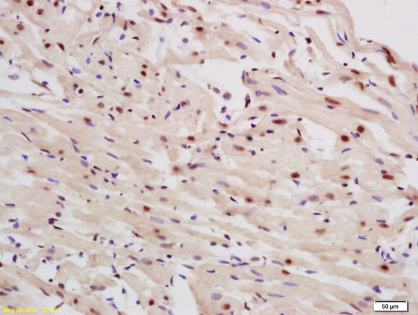
Formalin-fixed and paraffin embedded rat heart labeled with Rabbit Anti P63 protein/P51A Polyclonal Antibody, Unconjugated (bs-0723R) at 1:200 followed by conjugation to the secondary antibody and DAB staining
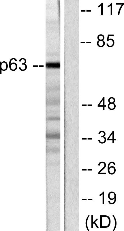
 at 1:1000 overnight at 4°C followed by a conjugated secondary antibody for 60 minutes at 37°C.")
Formalin-fixed and paraffin embedded rat heart labeled with Rabbit Anti P63 protein/P51A Polyclonal Antibody, Unconjugated (bs-0723R) at 1:200 followed by conjugation to the secondary antibody and DAB staining
p63 Polyclonal Antibody
BS-0723R
ApplicationsImmunoFluorescence, Western Blot, ELISA, ImmunoCytoChemistry, ImmunoHistoChemistry, ImmunoHistoChemistry Frozen, ImmunoHistoChemistry Paraffin
Product group Antibodies
ReactivityBovine, Canine, Equine, Guinea Pig, Human, Mouse, Porcine, Rabbit, Rat, Sheep
TargetTP63
Overview
- SupplierBioss
- Product Namep63 Polyclonal Antibody
- Delivery Days Customer16
- ApplicationsImmunoFluorescence, Western Blot, ELISA, ImmunoCytoChemistry, ImmunoHistoChemistry, ImmunoHistoChemistry Frozen, ImmunoHistoChemistry Paraffin
- Applications SupplierWB(1:300-5000), ELISA(1:500-1000), IHC-P(1:200-400), IHC-F(1:100-500), IF(IHC-P)(1:50-200), IF(IHC-F)(1:50-200), IF(ICC)(1:50-200)
- CertificationResearch Use Only
- ClonalityPolyclonal
- Concentration1 ug/ul
- ConjugateUnconjugated
- Gene ID8626
- Target nameTP63
- Target descriptiontumor protein p63
- Target synonymsAIS, B(p51A), B(p51B), EEC3, KET, LMS, NBP, OFC8, RHS, SHFM4, TP53CP, TP53L, TP73L, p40, p51, p53CP, p63, p73H, p73L, tumor protein 63, amplified in squamous cell carcinoma, chronic ulcerative stomatitis protein, keratinocyte transcription factor KET, transformation-related protein 63, tumor protein p53-competing protein
- HostRabbit
- IsotypeIgG
- Protein IDQ9H3D4
- Protein NameTumor protein 63
- ReactivityBovine, Canine, Equine, Guinea Pig, Human, Mouse, Porcine, Rabbit, Rat, Sheep
- Storage Instruction-20°C
- UNSPSC41116161
References
- Influenza Virus Infects Epithelial Stem/Progenitor Cells of the Distal Lung: Impact on Fgfr2b-Driven Epithelial Repair. Quantius J et al., 2016 Jun, PLoS PathogRead this paper
- An improved method for the isolation and culture of rat epidermal stem cells. Yang RH et al., 2013, Int J Clin Exp PatholRead this paper
Datasheet
Related products
Product group Antibodies
Anti-p63 AntibodyA96644
ApplicationsImmunoFluorescence, Western Blot, ELISA
ReactivityHuman, Mouse, Rat
- SizePrice
Product group Antibodies
Anti-TP63 Antibody144-12937
ApplicationsImmunoFluorescence, Western Blot
ReactivityHuman, Mouse, Rat
TargetTP63
- SizePrice


Product group Antibodies
Anti-TP63 AntibodyAMAB91224
ApplicationsWestern Blot, ImmunoHistoChemistry
ReactivityHuman
TargetTP63
- SizePrice
Product group Antibodies

Anti-p63 [BU5]Ab00871-10.0
ApplicationsFlow Cytometry, ImmunoFluorescence
ReactivityHuman
TargetTP63
- SizePrice
Product group Antibodies

TP63 / p63 AntibodyLS-C830518
ApplicationsELISA, ImmunoHistoChemistry
ReactivityHuman, Mouse, Rat
TargetTP63
- SizePrice
Product group Antibodies

ApplicationsWestern Blot, ELISA
ReactivityBovine, Canine, Human, Mouse, Porcine, Rat
TargetTP63
- SizePrice
Product group Antibodies
ApplicationsImmunoPrecipitation, Western Blot, ImmunoCytoChemistry, ImmunoHistoChemistry
TargetTP63
- SizePrice
Product group Antibodies
TP63 AntibodyCSB-PA005081
ApplicationsWestern Blot, ELISA
ReactivityHuman, Mouse, Rat
TargetTP63
- SizePrice