

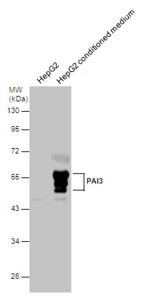
PAI3 antibody detects PAI3 protein by western blot analysis. HepG2 whole cell extracts (30 μg) and conditioned medium were separated by 10% SDS-PAGE, and the membrane was blotted with PAI3 antibody (GTX113795) diluted at 1:10000.
 dilution: 1:500.
Antigen Retrieval: Trilogy? (EDTA based, pH 8.0) buffer, 15min")
 antibody at 1:200 dilution.")
PAI3 antibody detects PAI3 protein by western blot analysis. HepG2 whole cell extracts (30 μg) and conditioned medium were separated by 10% SDS-PAGE, and the membrane was blotted with PAI3 antibody (GTX113795) diluted at 1:10000.
PAI3 antibody
GTX113795
ApplicationsImmunoFluorescence, Western Blot, ImmunoCytoChemistry, ImmunoHistoChemistry, ImmunoHistoChemistry Paraffin
Product group Antibodies
ReactivityHuman
TargetSERPINA5
Overview
- SupplierGeneTex
- Product NamePAI3 antibody
- Delivery Days Customer9
- Application Supplier NoteWB: 1:500-1:10000. ICC/IF: 1:100-1:1000. IHC-P: 1:100-1:1000. *Optimal dilutions/concentrations should be determined by the researcher.Not tested in other applications.
- ApplicationsImmunoFluorescence, Western Blot, ImmunoCytoChemistry, ImmunoHistoChemistry, ImmunoHistoChemistry Paraffin
- CertificationResearch Use Only
- ClonalityPolyclonal
- Concentration1 mg/ml
- ConjugateUnconjugated
- Gene ID5104
- Target nameSERPINA5
- Target descriptionserpin family A member 5
- Target synonymsPAI-3, PAI3, PCI, PCI-B, PLANH3, PROCI, plasma serine protease inhibitor, acrosomal serine protease inhibitor, plasminogen activator inhibitor III, plasminogen activator inhibitor-3, protein C inhibitor, serine (or cysteine) proteinase inhibitor, clade A (alpha-1 antiproteinase, antitrypsin), member 5, serpin peptidase inhibitor, clade A (alpha-1 antiproteinase, antitrypsin), member 5
- HostRabbit
- IsotypeIgG
- Protein IDP05154
- Protein NamePlasma serine protease inhibitor
- ReactivityHuman
- Storage Instruction-20°C or -80°C,2°C to 8°C
- UNSPSC41116161
Datasheet
Related products
Product group Antibodies
SERPINA5 AntibodyCSB-PA003700
ApplicationsWestern Blot, ELISA, ImmunoHistoChemistry
ReactivityHuman
TargetSERPINA5
- SizePrice
Product group Antibodies
Anti-Protein C inhibitor/SERPINA5 Antibody Picoband(r)A01916-1-CARRIER-FREE
ApplicationsWestern Blot, ImmunoHistoChemistry
ReactivityHuman, Mouse, Rat
TargetSERPINA5
- SizePrice
Product group Antibodies

PAI-3 AntibodyABX019149
ApplicationsImmunoFluorescence, Western Blot, ImmunoCytoChemistry
- SizePrice
Product group Antibodies
ApplicationsWestern Blot, ImmunoCytoChemistry
ReactivityHuman
- SizePrice

Product group Antibodies
Anti-SERPINA5 AntibodyHPA078846
ApplicationsImmunoHistoChemistry
ReactivityHuman
TargetSERPINA5
- SizePrice
Product group Antibodies

SERPINA5 / PCI AntibodyLS-C401126
ApplicationsWestern Blot, ELISA
ReactivityHuman
TargetSERPINA5
- SizePrice
Product group Antibodies
Serpina5 Polyclonal AntibodyCAC11722
ApplicationsImmunoFluorescence, Western Blot, ELISA, ImmunoHistoChemistry
ReactivityMouse, Rat
TargetSERPINA5
- SizePrice
![PAI3 antibody [N3C3] detects PAI3 protein by western blot analysis. HepG2 whole cell extracts and HepG2 conditioned medium (30 μg) were separated by 10% SDS-PAGE, and the membrane was blotted with PAI3 antibody [N3C3] (GTX106655) diluted at 1:5000.](https://www.genetex.com/upload/website/prouct_img/normal/GTX106655/GTX106655_40135_20151015_WB_w_23060120_729.webp)
Product group Antibodies
PAI3 antibody [N3C3]GTX106655
ApplicationsWestern Blot, ImmunoHistoChemistry, ImmunoHistoChemistry Paraffin
ReactivityHuman
TargetSERPINA5
- SizePrice
Product group Antibodies
PAI3 antibody [11B7]GTX52995
ApplicationsWestern Blot
ReactivityHuman
TargetSERPINA5
- SizePrice
Product group Antibodies
Anti-SERPINA5 (N-term) Antibody102-22165
ApplicationsWestern Blot
TargetSERPINA5
- SizePrice