

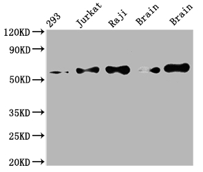
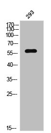
Western Blot Positive WB detected in: 293 whole cell lysate, Jurkat whole cell lysate, Raji whole cell lysate, Mouse brain tissue, Rat brain tissue All lanes: PAK2 antibody at 1:2000 Secondary Goat polyclonal to rabbit IgG at 1/50000 dilution Predicted band size: 59 kDa Observed band size: 59 kDa
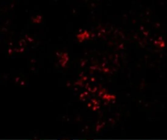
 at 1:50. The cells were fixed with 70% Ethylalcohol (18h) and then incubated in 10% normal goat serum to block non-specific protein-protein interactions followedby the antibody (1microg/1*106 cells) for 1 h at 4°C.The secondary antibody used was FITC-conjugated goat anti-rabbit IgG (H+L) at 1/200 dilution for 30min at 4°C. Control antibody (green line) was Rabbit IgG (1microg/1*106 cells) used under the same conditions. Acquisition of >10,000 events was performed.")
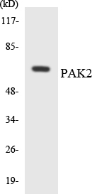
 Lane 2: CSB-RA592787A0HU(2microg)+ Raji whole cell lysate(500microg) Lane 3: Raji whole cell lysate (10microg)")
Western Blot Positive WB detected in: 293 whole cell lysate, Jurkat whole cell lysate, Raji whole cell lysate, Mouse brain tissue, Rat brain tissue All lanes: PAK2 antibody at 1:2000 Secondary Goat polyclonal to rabbit IgG at 1/50000 dilution Predicted band size: 59 kDa Observed band size: 59 kDa
PAK2 Recombinant Monoclonal Antibody
CSB-RA592787A0HU
ApplicationsFlow Cytometry, ImmunoPrecipitation, Western Blot, ELISA
Product group Antibodies
ReactivityHuman, Mouse, Rat
TargetPAK2
Overview
- SupplierCusabio
- Product NamePAK2 Recombinant Monoclonal Antibody
- Delivery Days Customer20
- ApplicationsFlow Cytometry, ImmunoPrecipitation, Western Blot, ELISA
- CertificationResearch Use Only
- ClonalityMonoclonal
- Clone ID6D12
- ConjugateUnconjugated
- Gene ID5062
- Target namePAK2
- Target descriptionp21 (RAC1) activated kinase 2
- Target synonymsKNO2, PAK65, PAKgamma, serine/threonine-protein kinase PAK 2, PAK-2, S6/H4 kinase, gamma-PAK, p21 (CDKN1A)-activated kinase 2, p21 protein (Cdc42/Rac)-activated kinase 2, p21-activated kinase 2, p58
- IsotypeIgG
- Protein IDQ13177
- Protein NameSerine/threonine-protein kinase PAK 2
- Scientific DescriptionSerine/threonine protein kinase that plays a role in a variety of different signaling pathways including cytoskeleton regulation, cell motility, cell cycle progression, apoptosis or proliferation. Acts as downstream effector of the small GTPases CDC42 and RAC1. Activation by the binding of active CDC42 and RAC1 results in a conformational change and a subsequent autophosphorylation on several serine and/or threonine residues. Full-length PAK2 stimulates cell survival and cell growth. Phosphorylates MAPK4 and MAPK6 and activates the downstream target MAPKAPK5, a regulator of F-actin polymerization and cell migration. Phosphorylates JUN and plays an important role in EGF-induced cell proliferation. Phosphorylates many other substrates including histone H4 to promote assembly of H3.3 and H4 into nucleosomes, BAD, ribosomal protein S6, or MBP. Additionally, associates with ARHGEF7 and GIT1 to perform kinase-independent functions such as spindle orientation control during mitosis. On the other hand, apoptotic stimuli such as DNA damage lead to caspase-mediated cleavage of PAK2, generating PAK-2p34, an active p34 fragment that translocates to the nucleus and promotes cellular apoptosis involving the JNK signaling pathway. Caspase-activated PAK2 phosphorylates MKNK1 and reduces cellular translation.
- ReactivityHuman, Mouse, Rat
- Storage Instruction-20°C or -80°C
- UNSPSC41116161
Related products
Product group Antibodies
Anti-PAK2 Antibody144-07333
ApplicationsImmunoFluorescence, Western Blot, ImmunoHistoChemistry
ReactivityHuman, Mouse, Rat
TargetPAK2
- SizePrice
Product group Antibodies
ApplicationsWestern Blot, ImmunoHistoChemistry
ReactivityMouse
TargetPAK2
- SizePrice
Product group Antibodies
PAK2 Recombinant Antibody, Biotin ConjugatedBSM-61673R-BIOTIN
ApplicationsWestern Blot, ImmunoHistoChemistry, ImmunoHistoChemistry Frozen, ImmunoHistoChemistry Paraffin
ReactivityHuman, Mouse, Rat
TargetPAK2
- SizePrice
Product group Antibodies
Phospho-PAK2 (S20) AntibodyCSB-PA010536
ApplicationsWestern Blot, ELISA, ImmunoHistoChemistry
ReactivityHuman, Mouse, Rat
TargetPAK2
- SizePrice
Product group Antibodies
Anti-PAK2 AntibodyA96147
ApplicationsWestern Blot, ELISA, ImmunoHistoChemistry
ReactivityHuman, Mouse, Rat
- SizePrice
Product group Antibodies
PAK2 antibodyGTX31742
ApplicationsWestern Blot, ELISA, ImmunoHistoChemistry, ImmunoHistoChemistry Paraffin
ReactivityHuman, Mouse, Rat
TargetPAK2
- SizePrice
Product group Antibodies

PAK2 AntibodyLS-C400945
ApplicationsELISA, ImmunoHistoChemistry
ReactivityHuman, Mouse, Rat
TargetPAK2
- SizePrice

Product group Antibodies
Anti-PAK2 Antibody Picoband(r)A01419-4-CARRIER-FREE
ApplicationsFlow Cytometry, ImmunoFluorescence, Western Blot, ELISA, ImmunoCytoChemistry, ImmunoHistoChemistry
ReactivityHuman, Mouse, Rat
TargetPAK2
- SizePrice