

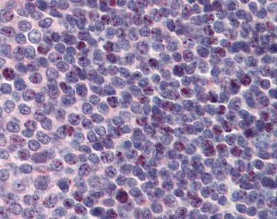
IHC-P analysis of human spleen tissue using GTX47349 PCBP1 antibody at 5μg/ml.


IHC-P analysis of human spleen tissue using GTX47349 PCBP1 antibody at 5μg/ml.
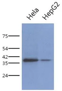
PCBP1 antibody, Internal
GTX47349
ApplicationsWestern Blot, ImmunoHistoChemistry, ImmunoHistoChemistry Paraffin
Product group Antibodies
ReactivityHuman
TargetPCBP1
Overview
- SupplierGeneTex
- Product NamePCBP1 antibody, Internal
- Delivery Days Customer9
- Application Supplier NoteWB: 0.2-2.5 ug/ml. IHC-P: 2-10 ug/ml. *Optimal dilutions/concentrations should be determined by the researcher.Not tested in other applications.
- ApplicationsWestern Blot, ImmunoHistoChemistry, ImmunoHistoChemistry Paraffin
- CertificationResearch Use Only
- ClonalityPolyclonal
- Concentration0.5-1 mg/ml
- ConjugateUnconjugated
- Gene ID5093
- Target namePCBP1
- Target descriptionpoly(rC) binding protein 1
- Target synonymsHEL-S-85, HNRPE1, HNRPX, hnRNP-E1, hnRNP-X, poly(rC)-binding protein 1, alpha-CP1, epididymis secretory protein Li 85, heterogeneous nuclear ribonucleoprotein E1, heterogenous nuclear ribonucleoprotein E1, heterogenous nuclear ribonucleoprotein X, nucleic acid-binding protein SUB2.3
- HostRabbit
- IsotypeIgG
- Protein IDQ15365
- Protein NamePoly(rC)-binding protein 1
- Scientific DescriptionThis intronless gene is thought to have been generated by retrotransposition of a fully processed PCBP-2 mRNA. This gene and PCBP-2 have paralogues (PCBP3 and PCBP4) which are thought to have arisen as a result of duplication events of entire genes. The protein encoded by this gene appears to be multifunctional. It along with PCBP-2 and hnRNPK corresponds to the major cellular poly(rC)-binding protein. It contains three K-homologous (KH) domains which may be involved in RNA binding. This encoded protein together with PCBP-2 also functions as translational coactivators of poliovirus RNA via a sequence-specific interaction with stem-loop IV of the IRES and promote poliovirus RNA replication by binding to its 5-terminal cloverleaf structure. It has also been implicated in translational control of the 15-lipoxygenase mRNA, human Papillomavirus type 16 L2 mRNA, and hepatitis A virus RNA. The encoded protein is also suggested to play a part in formation of a sequence-specific alpha-globin mRNP complex which is associated with alpha-globin mRNA stability. [provided by RefSeq, Jul 2008]
- ReactivityHuman
- Storage Instruction-20°C or -80°C,2°C to 8°C
- UNSPSC12352203
Datasheet
Related products
Product group Antibodies
Anti-PCBP1 Antibody144-01044
ApplicationsImmunoFluorescence, Western Blot, ImmunoHistoChemistry
ReactivityHuman, Mouse, Rat
TargetPCBP1
- SizePrice
Product group Antibodies
PCBP1 Recombinant AntibodyBSM-62413R
ApplicationsImmunoPrecipitation, Western Blot
ReactivityHuman, Mouse, Rat
TargetPCBP1
- SizePrice
Product group Antibodies
Anti-hnRNP E1 AntibodyA28251
ApplicationsWestern Blot
ReactivityHuman, Mouse, Rat
- SizePrice
Product group Antibodies
PCBP1 antibody, InternalGTX47350
ApplicationsWestern Blot
ReactivityHuman
TargetPCBP1
- SizePrice
Product group Antibodies
PCBP1 antibodyGTX64891
ApplicationsImmunoFluorescence, Western Blot, ImmunoCytoChemistry, ImmunoHistoChemistry, ImmunoHistoChemistry Paraffin
ReactivityHuman, Mouse, Rat
TargetPCBP1
- SizePrice

Product group Antibodies
Anti-PCBP1 AntibodyHPA043803
ApplicationsWestern Blot, ImmunoCytoChemistry
ReactivityHuman
TargetPCBP1
- SizePrice
Product group Antibodies
PCBP1 antibody [AT2A10]GTX53759
ApplicationsWestern Blot, ELISA
ReactivityHuman
TargetPCBP1
- SizePrice
Product group Antibodies
PCBP1 AntibodyCSB-PA613590LA01HU
ApplicationsELISA
ReactivityHuman
TargetPCBP1
- SizePrice
Product group Antibodies
References

ApplicationsWestern Blot, ELISA
ReactivityCanine, Human, Mouse, Rat
TargetPCBP1
- SizePrice