

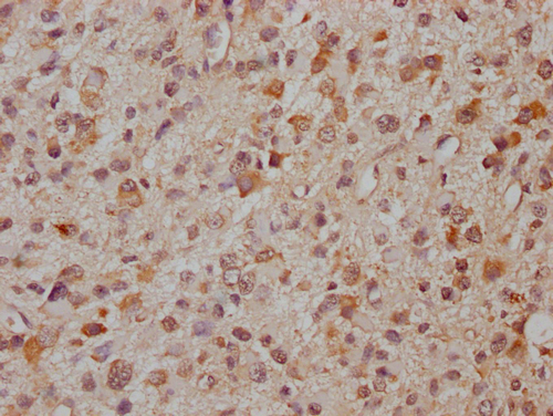
PCDHA8 Antibody (aa773-800, FITC)
LS-C238775
ApplicationsImmunoFluorescence, Western Blot, ELISA, ImmunoCytoChemistry, ImmunoHistoChemistry
Product group Antibodies
TargetPCDHA8
Overview
- SupplierLifeSpan BioSciences
- Product NamePCDHA8 Antibody (aa773-800, FITC)
- Delivery Days Customer23
- Application Supplier NoteThe applications listed have been tested for the unconjugated form of this product. Other forms have not been tested.
- ApplicationsImmunoFluorescence, Western Blot, ELISA, ImmunoCytoChemistry, ImmunoHistoChemistry
- Applications SupplierELISA, ICC, IF, IHC, WB The applications listed have been tested for the unconjugated form of this product. Other forms have not been tested.
- CertificationResearch Use Only
- ClonalityPolyclonal
- ConjugateFITC
- Estimated Purity...
- Gene ID56140
- Target namePCDHA8
- Target descriptionprotocadherin alpha 8
- Target synonymsKIAA0345-like 6; PCDH-ALPHA8; PCDH-alpha-8; protocadherin alpha-8
- HostRabbit
- IsotypeIgG
- Storage Instruction-20°C,2°C to 8°C
- UNSPSC12352203
Related products
Product group Antibodies

PCDHA8 Antibody (aa773-800)LS-C163423
ApplicationsImmunoFluorescence, Western Blot, ImmunoHistoChemistry, ImmunoHistoChemistry Paraffin
TargetPCDHA8
- SizePrice
Product group Antibodies
PCDHA8 AntibodyCSB-PA897294LA01HU
ApplicationsELISA, ImmunoHistoChemistry
ReactivityHuman
TargetPCDHA8
- SizePrice

Product group Antibodies
Anti-PCDHA8 AntibodyHPA044585
ApplicationsImmunoHistoChemistry
ReactivityHuman
TargetPCDHA8
- SizePrice

Product group Antibodies
Anti-PCDHA8 Antibody Picoband(r)A15533-CARRIER-FREE
ApplicationsFlow Cytometry, Western Blot, ELISA
TargetPCDHA8
- SizePrice
Product group Antibodies
PCDHA8 AntibodyPACO64893
ApplicationsELISA, ImmunoHistoChemistry
TargetPCDHA8
- SizePrice