

PD-L1 antibody [HL1056]
GTX636033
ApplicationsImmunoFluorescence, Western Blot, ImmunoCytoChemistry
Product group Antibodies
ReactivityHuman
TargetCD274
Overview
- SupplierGeneTex
- Product NamePD-L1 antibody [HL1056]
- Delivery Days Customer9
- Application Supplier NoteWB: 1:500-1:3000. *Optimal dilutions/concentrations should be determined by the researcher.Not tested in other applications.
- ApplicationsImmunoFluorescence, Western Blot, ImmunoCytoChemistry
- CertificationResearch Use Only
- ClonalityMonoclonal
- Clone IDHL1056
- Concentration1 mg/ml
- ConjugateUnconjugated
- Gene ID29126
- Target nameCD274
- Target descriptionCD274 molecule
- Target synonymsADMIO5, B7-H, B7H1, PD-L1, PDCD1L1, PDCD1LG1, PDL1, hPD-L1, programmed cell death 1 ligand 1, B7 homolog 1, CD274 antigen, PDCD1 ligand 1
- HostRabbit
- IsotypeIgG
- Protein IDQ9NZQ7
- Protein NameProgrammed cell death 1 ligand 1
- Scientific DescriptionThis gene encodes an immune inhibitory receptor ligand that is expressed by hematopoietic and non-hematopoietic cells, such as T cells and B cells and various types of tumor cells. The encoded protein is a type I transmembrane protein that has immunoglobulin V-like and C-like domains. Interaction of this ligand with its receptor inhibits T-cell activation and cytokine production. During infection or inflammation of normal tissue, this interaction is important for preventing autoimmunity by maintaining homeostasis of the immune response. In tumor microenvironments, this interaction provides an immune escape for tumor cells through cytotoxic T-cell inactivation. Expression of this gene in tumor cells is considered to be prognostic in many types of human malignancies, including colon cancer and renal cell carcinoma. Alternative splicing results in multiple transcript variants. [provided by RefSeq, Sep 2015]
- ReactivityHuman
- Storage Instruction-20°C or -80°C,2°C to 8°C
- UNSPSC12352203
Datasheet
Related products
Product group Antibodies
CD274 AntibodyCSB-PA878942ESR1HU
ApplicationsELISA, ImmunoHistoChemistry
ReactivityHuman
TargetCD274
- SizePrice
Product group Antibodies
PD-L1 Polyclonal AntibodyBS-22022R
ApplicationsImmunoFluorescence, Western Blot, ELISA, ImmunoCytoChemistry, ImmunoHistoChemistry, ImmunoHistoChemistry Frozen, ImmunoHistoChemistry Paraffin
ReactivityHuman, Mouse, Rat
TargetCD274
- SizePrice
Product group Antibodies
ApplicationsFlow Cytometry
TargetCD274
- SizePrice
Product group Antibodies
References
PD-L1 antibody, C-termGTX89590
ApplicationsFlow Cytometry, ImmunoFluorescence, Western Blot, ImmunoCytoChemistry
ReactivityHuman
TargetCD274
- SizePrice
Product group Antibodies
PD-L1 antibody [9E5]GTX52553
ApplicationsWestern Blot
ReactivityHuman
TargetCD274
- SizePrice
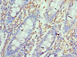
![IHC-P analysis of human lung tissue using GTX57193 PD-L1 antibody [IHC411]](https://www.genetex.com/upload/website/prouct_img/normal/GTX57193/GTX57193_20180619_IHC-P_w_23061123_830.webp)
Product group Antibodies
PD-L1 antibody [IHC411]GTX57193
ApplicationsImmunoFluorescence, ImmunoCytoChemistry, ImmunoHistoChemistry, ImmunoHistoChemistry Paraffin
ReactivityHuman
TargetCD274
- SizePrice
Product group Antibodies
PD-L1 antibody [H302] HistoMAX(tm)GTX639925
ApplicationsImmunoHistoChemistry, ImmunoHistoChemistry Paraffin
ReactivityHuman
TargetCD274
- SizePrice
Product group Antibodies
PD-L1 antibody [HL2985]GTX640395
ApplicationsFlow Cytometry, ImmunoFluorescence, Western Blot, ImmunoCytoChemistry, ImmunoHistoChemistry, ImmunoHistoChemistry Paraffin
ReactivityHuman
TargetCD274
- SizePrice
Product group Antibodies

ApplicationsWestern Blot, ELISA
ReactivityHuman
TargetCD274
- SizePrice
Product group Antibodies
References
PD-L1 antibody [HL1041]GTX635975
ApplicationsFlow Cytometry, ImmunoFluorescence, Western Blot, ImmunoCytoChemistry, ImmunoHistoChemistry, ImmunoHistoChemistry Paraffin
ReactivityHuman
TargetCD274
- SizePrice