

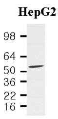
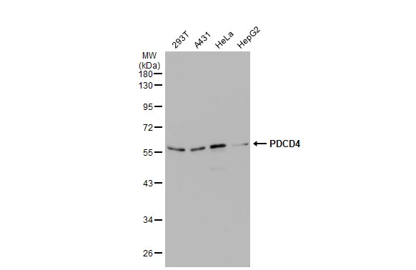
Cell lysates of HepG2(40ug) were resolved by SDS-PAGE, transferred to NC membrane and probed with anti-human PDCD4 (1:2,000). Proteins were visualized using a goat anti-mouse secondary antibody conjugated to HRP and an ECL detection system.
Cell lysates of HepG2(40ug) were resolved by SDS-PAGE, transferred to NC membrane and probed with anti-human PDCD4 (1:2,000). Proteins were visualized using a goat anti-mouse secondary antibody conjugated to HRP and an ECL detection system.
PDCD4 antibody [k4C1]
GTX50048
ApplicationsWestern Blot, ELISA
Product group Antibodies
ReactivityHuman
TargetPDCD4
Overview
- SupplierGeneTex
- Product NamePDCD4 antibody [k4C1]
- Delivery Days Customer9
- Application Supplier NoteWe recommend the following starting dilutions:Western Blot: Use at 1:1,000~ 3,000.Optimal working concentrations should be determined experimentally by the end user.
- ApplicationsWestern Blot, ELISA
- CertificationResearch Use Only
- ClonalityMonoclonal
- Clone IDk4C1
- Concentration1 mg/ml
- ConjugateUnconjugated
- Gene ID27250
- Target namePDCD4
- Target descriptionprogrammed cell death 4
- Target synonymsH731, programmed cell death protein 4, neoplastic transformation inhibitor protein, nuclear antigen H731, programmed cell death 4 (neoplastic transformation inhibitor), protein 197/15a
- HostMouse
- IsotypeIgG1
- Protein IDQ53EL6
- Protein NameProgrammed cell death protein 4
- Scientific DescriptionThis gene is a tumor suppressor and encodes a protein that binds to the eukaryotic translation initiation factor 4A1 and inhibits its function by preventing RNA binding. Alternative splicing results in multiple transcript variants. [provided by RefSeq, Dec 2010]
- ReactivityHuman
- Storage Instruction-20°C or -80°C,2°C to 8°C
- UNSPSC41116161
Datasheet
Related products
Product group Antibodies
PDCD4 AntibodyCSB-PA003720
ApplicationsImmunoFluorescence, Western Blot, ELISA, ImmunoHistoChemistry
ReactivityHuman, Mouse, Rat
TargetPDCD4
- SizePrice
Product group Antibodies
Anti-PDCD4 AntibodyA83067
ApplicationsWestern Blot, ELISA
ReactivityHuman, Mouse
- SizePrice

Product group Antibodies
Anti-PDCD4 Antibody Picoband(r)A01105-3-CARRIER-FREE
ApplicationsFlow Cytometry, Western Blot
ReactivityHuman
TargetPDCD4
- SizePrice
Product group Antibodies

PDCD4 AntibodyLS-C831489
ApplicationsWestern Blot, ImmunoHistoChemistry
ReactivityHuman, Mouse, Rat
TargetPDCD4
- SizePrice

Product group Antibodies
Anti-PDCD4 AntibodyHPA001032
ApplicationsWestern Blot, ChIP Chromatin ImmunoPrecipitation, ImmunoHistoChemistry
ReactivityHuman, Mouse, Rat
TargetPDCD4
- SizePrice
Product group Antibodies
PDCD4 Polyclonal AntibodyBS-1608R
ApplicationsImmunoFluorescence, Western Blot, ELISA, ImmunoCytoChemistry, ImmunoHistoChemistry, ImmunoHistoChemistry Frozen, ImmunoHistoChemistry Paraffin
ReactivityHuman, Mouse, Rat
TargetPDCD4
- SizePrice
Product group Antibodies
PDCD4 antibodyGTX104901
ApplicationsImmunoFluorescence, Western Blot, ImmunoCytoChemistry
ReactivityHuman, Mouse
TargetPDCD4
- SizePrice
Product group Antibodies
ApplicationsWestern Blot, ELISA, ImmunoHistoChemistry, Other Application
ReactivityHuman, Mouse, Rat
TargetPDCD4
- SizePrice