

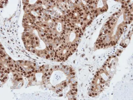
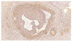
Immunohistochemical analysis of paraffin-embedded human endo mitral, using PDE4C(GTX106268) antibody at 1:100 dilution.
Antigen Retrieval: Trilogy? (EDTA based, pH 8.0) buffer, 15min
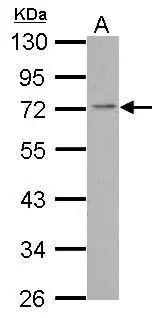
![Mouse tissue extract (50 μg) was separated by 7.5% SDS-PAGE, and the membrane was blotted with PDE4C antibody [N3C3] (GTX106268) diluted at 1:1000. The HRP-conjugated anti-rabbit IgG antibody (GTX213110-01) was used to detect the primary antibody.](https://www.genetex.com/upload/website/prouct_img/normal/GTX106268/GTX106268_39890_20220304_WB_M_lung_w_23060120_608.webp "Mouse tissue extract (50 μg) was separated by 7.5% SDS-PAGE, and the membrane was blotted with PDE4C antibody [N3C3] (GTX106268) diluted at 1:1000. The HRP-conjugated anti-rabbit IgG antibody (GTX213110-01) was used to detect the primary antibody.")
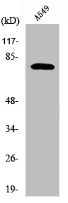
 A:293T 7.5% SDS PAGE GTX106268 diluted at 1:1000")
Immunohistochemical analysis of paraffin-embedded human endo mitral, using PDE4C(GTX106268) antibody at 1:100 dilution.
Antigen Retrieval: Trilogy? (EDTA based, pH 8.0) buffer, 15min
PDE4C antibody [N3C3]
GTX106268
ApplicationsWestern Blot, ImmunoHistoChemistry, ImmunoHistoChemistry Paraffin
Product group Antibodies
ReactivityHuman, Mouse
TargetPDE4C
Overview
- SupplierGeneTex
- Product NamePDE4C antibody [N3C3]
- Delivery Days Customer9
- Application Supplier NoteWB: 1:500-1:3000. IHC-P: 1:100-1:1000. *Optimal dilutions/concentrations should be determined by the researcher.Not tested in other applications.
- ApplicationsWestern Blot, ImmunoHistoChemistry, ImmunoHistoChemistry Paraffin
- CertificationResearch Use Only
- ClonalityPolyclonal
- Concentration0.67 mg/ml
- ConjugateUnconjugated
- Gene ID5143
- Target namePDE4C
- Target descriptionphosphodiesterase 4C
- Target synonymsDPDE1, PDE21, 3',5'-cyclic-AMP phosphodiesterase 4C, cAMP-specific 3',5'-cyclic phosphodiesterase 4C, cAMP-specific phosphodiesterase 4C, dunce-like phosphodiesterase E1, phosphodiesterase 4C, cAMP-specific (phosphodiesterase E1 dunce homolog, Drosophila)
- HostRabbit
- IsotypeIgG
- Protein IDQ08493
- Protein Name3',5'-cyclic-AMP phosphodiesterase 4C
- Scientific DescriptionCyclic nucleotides are important second messengers that regulate and mediate a number of cellular responses to extracellular signals, such as hormones, light, and neurotransmitters. Cyclic nucleotide phosphodiesterases (PDEs) regulate the cellular concentrations of cyclic nucleotides and thereby play a role in signal transduction. PDE4C is a class IV cAMP-specific PDE (summary by Milatovich et al., 1994 [PubMed 8009369]).[supplied by OMIM]
- ReactivityHuman, Mouse
- Storage Instruction-20°C or -80°C,2°C to 8°C
- UNSPSC12352203
Datasheet
Related products

Product group Antibodies
Anti-PDE4C AntibodyA07630-1
ApplicationsWestern Blot, ELISA, ImmunoHistoChemistry
ReactivityHuman, Mouse, Rat
TargetPDE4C
- SizePrice
Product group Antibodies
Anti-PDE4C Antibody144-62602
ApplicationsWestern Blot
ReactivityHuman, Rat
TargetPDE4C
- SizePrice

Product group Antibodies
Anti-PDE4C AntibodyHPA048975
ApplicationsImmunoHistoChemistry
ReactivityHuman
TargetPDE4C
- SizePrice
Product group Antibodies
PDE4C AntibodyCSB-PA832211
ApplicationsWestern Blot, ELISA
ReactivityHuman, Mouse, Rat
TargetPDE4C
- SizePrice
Product group Antibodies
PDE4C antibodyGTX14608
ApplicationsImmunoFluorescence, ImmunoPrecipitation, Western Blot, ELISA, ImmunoCytoChemistry, ImmunoHistoChemistry, ImmunoHistoChemistry Frozen, ImmunoHistoChemistry Paraffin
ReactivityHuman, Mouse, Rat
TargetPDE4C
- SizePrice
Product group Antibodies
PDE4C antibody [N3C2], InternalGTX118307
ApplicationsWestern Blot
ReactivityHuman
TargetPDE4C
- SizePrice
Product group Antibodies

PDE4C AntibodyLS-C748099
ApplicationsWestern Blot
ReactivityHuman, Rat
TargetPDE4C
- SizePrice

Product group Antibodies
Anti-PDE4C AntibodyA97357
ApplicationsWestern Blot, ELISA
ReactivityHuman, Mouse, Rat
- SizePrice