

PDGF-BB antibody
GTX10845
ApplicationsWestern Blot, ELISA, Neutralisation/Blocking
Product group Antibodies
ReactivityHuman
TargetPDGFB
Overview
- SupplierGeneTex
- Product NamePDGF-BB antibody
- Delivery Days Customer9
- ApplicationsWestern Blot, ELISA, Neutralisation/Blocking
- CertificationResearch Use Only
- ClonalityPolyclonal
- ConjugateUnconjugated
- Gene ID5155
- Target namePDGFB
- Target descriptionplatelet derived growth factor subunit B
- Target synonymsIBGC5, PDGF-2, PDGF2, SIS, SSV, c-sis, platelet-derived growth factor subunit B, PDGF subunit B, PDGF, B chain, becaplermin, epididymis secretory sperm binding protein, platelet-derived growth factor 2, platelet-derived growth factor B chain, platelet-derived growth factor beta polypeptide (simian sarcoma viral (v-sis) oncogene homolog), platelet-derived growth factor, beta polypeptide (oncogene SIS), proto-oncogene c-Sis
- HostGoat
- IsotypeIgG
- Protein IDP01127
- Protein NamePlatelet-derived growth factor subunit B
- Scientific DescriptionPlatelet-Derived Growth Factor (PDGF) in serum is the principal mitogen present for cells of mesenchymal origin. PDGF is localized in alpha-granules of platelets and released during clot formation. PDGF from human platelets has been purified and described as a cationic glycoprotein (pI 9.5 to 10.4) having a molecular weight of approximately 30 kD and composed of two covalently linked subunits, designated as chains A (16 kD) and B (14 kD). In platelets, approximately 70% of the PDGF is present as the AB dimer, with most of the remainder as BB.Purified human PDGF shows substantial size heterogeneity, ranging from 27 to 31 kD, probably due to the presence of isoforms, glycosylation processing, aging of the platelets, and partial proteolysis during purification. The A and B chains are 40% homologous in sequence and are encoded by distinctly different genes. Each chain contains 8 cysteine residues, which are involved in intra- and inter-chain disulfide bonds. Cleavage of these bonds by reduction causes irreversible loss of biological activity. PDGF elicits multifunctional actions with a variety of cells.
- ReactivityHuman
- Storage Instruction-20°C or -80°C,2°C to 8°C
- UNSPSC41116161
Datasheet
Related products
Product group Antibodies
Anti-PDGFB AntibodyA97356
ApplicationsWestern Blot, ELISA
ReactivityHuman, Mouse, Rat
- SizePrice
Product group Antibodies
Anti-PDGFB Antibody144-01195
ApplicationsWestern Blot, ImmunoHistoChemistry
ReactivityHuman, Mouse, Rat
TargetPDGFB
- SizePrice

Product group Antibodies
Anti-PDGF beta/PDGFB Antibody Picoband(r)A00348-CARRIER-FREE
ApplicationsWestern Blot
ReactivityHuman, Mouse, Rat
TargetPDGFB
- SizePrice
Product group Antibodies
References
PDGF-B Polyclonal AntibodyBS-0185R
ApplicationsFlow Cytometry, ImmunoFluorescence, Western Blot, ELISA, ImmunoCytoChemistry, ImmunoHistoChemistry, ImmunoHistoChemistry Frozen, ImmunoHistoChemistry Paraffin
ReactivityBovine, Canine, Equine, Human, Mouse, Rabbit, Rat, Sheep
TargetPDGFB
- SizePrice
Product group Antibodies

Goat anti-PDGFB precursorEB09553
ApplicationsFlow Cytometry, ImmunoFluorescence, ELISA
ReactivityBovine, Canine, Human, Mouse, Rat, Sheep
TargetPDGFB
- SizePrice
Product group Antibodies
ApplicationsImmunoPrecipitation, Western Blot, ImmunoCytoChemistry, ImmunoHistoChemistry
ReactivityPorcine
TargetPDGFB
- SizePrice
Product group Antibodies
PDGFB AntibodyCSB-PA020300
ApplicationsWestern Blot, ELISA, ImmunoHistoChemistry
ReactivityHuman, Mouse, Rat
TargetPDGFB
- SizePrice
Product group Antibodies

PDGF-BB Antibody (Preservative Free)LS-C343506
ApplicationsWestern Blot, ELISA
ReactivityHuman
TargetPDGFB
- SizePrice
![WB analysis of human placenta tissue lysates using GTX52467 PDGF beta antibody [5F66].](https://www.genetex.com/upload/website/prouct_img/normal/GTX52467/GTX52467_20220822_WB_22082302_220.webp)
Product group Antibodies
PDGF beta antibody [5F66]GTX52467
ApplicationsWestern Blot, ImmunoHistoChemistry, ImmunoHistoChemistry Paraffin
ReactivityHuman
TargetPDGFB
- SizePrice
Product group Antibodies
References
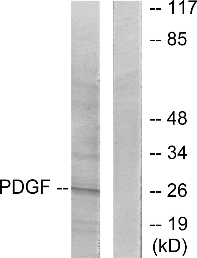
PDGF beta antibodyGTX54575
ApplicationsImmunoFluorescence, Western Blot, ImmunoCytoChemistry, ImmunoHistoChemistry, ImmunoHistoChemistry Paraffin
ReactivityHuman, Mouse, Rat
TargetPDGFB
- SizePrice