

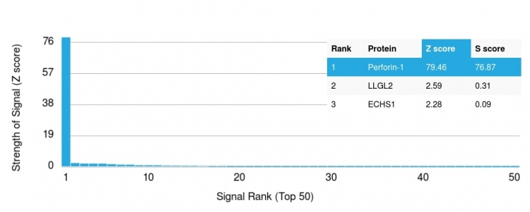
Analysis of Protein Array containing more than 19,000 full-length human proteins using Perforin-1 Mouse Monoclonal Antibody (PRF1/2468). Z- and S- Score: The Z-score represents the strength of a signal that a monoclonal antibody produces when binding to a particular protein on the HuProtTM array. Z-scores are described in units of standard deviations (SD’s) above the mean value of all signals generated on that array. If targets on HuProtTM are arranged in descending order of the Z-score, the S-score is the difference (also in units of SD’s) between the Z-score. S-score therefore represents the relative target specificity of a Monoclonal Antibody to its intended target. A Monoclonal Antibody is considered to specific to its intended target if the Monoclonal Antibody has an S-score of at least 2.5. For example, if a Monoclonal Antibody binds to protein X with a Z-score of 43 and to protein Y with a Z-score of 14, then the S-score for the binding of that Monoclonal Antibody to protein X is equal to 29.
Analysis of Protein Array containing more than 19,000 full-length human proteins using Perforin-1 Mouse Monoclonal Antibody (PRF1/2468). Z- and S- Score: The Z-score represents the strength of a signal that a monoclonal antibody produces when binding to a particular protein on the HuProtTM array. Z-scores are described in units of standard deviations (SD’s) above the mean value of all signals generated on that array. If targets on HuProtTM are arranged in descending order of the Z-score, the S-score is the difference (also in units of SD’s) between the Z-score. S-score therefore represents the relative target specificity of a Monoclonal Antibody to its intended target. A Monoclonal Antibody is considered to specific to its intended target if the Monoclonal Antibody has an S-score of at least 2.5. For example, if a Monoclonal Antibody binds to protein X with a Z-score of 43 and to protein Y with a Z-score of 14, then the S-score for the binding of that Monoclonal Antibody to protein X is equal to 29.
Perforin antibody [PRF1/2468]
GTX17985
ApplicationsELISA, Other Application
Product group Antibodies
ReactivityHuman
TargetPRF1
Overview
- SupplierGeneTex
- Product NamePerforin antibody [PRF1/2468]
- Delivery Days Customer9
- Application Supplier Note*Optimal dilutions/concentrations should be determined by the researcher.Not tested in other applications.
- ApplicationsELISA, Other Application
- CertificationResearch Use Only
- ClonalityMonoclonal
- Clone IDPRF1/2468
- Concentration0.2 mg/ml
- ConjugateUnconjugated
- Gene ID5551
- Target namePRF1
- Target descriptionperforin 1
- Target synonymsHPLH2, P1, PFP, perforin-1, cytolysin, lymphocyte pore-forming protein, perforin 1 (pore forming protein)
- HostMouse
- IsotypeIgG2b
- Protein IDP14222
- Protein NamePerforin-1
- Scientific DescriptionThis gene encodes a protein with structural similarities to complement component C9 that is important in immunity. This protein forms membrane pores that allow the release of granzymes and subsequent cytolysis of target cells. Whether pore formation occurs in the plasma membrane of target cells or in an endosomal membrane inside target cells is subject to debate. Mutations in this gene are associated with a variety of human disease including diabetes, multiple sclerosis, lymphomas, autoimmune lymphoproliferative syndrome (ALPS), aplastic anemia, and familial hemophagocytic lymphohistiocytosis type 2 (FHL2), a rare and lethal autosomal recessive disorder of early childhood. [provided by RefSeq, Aug 2017]
- ReactivityHuman
- Storage Instruction-20°C or -80°C,2°C to 8°C
- UNSPSC41116161
Datasheet
Related products
Product group Antibodies
Anti-PRF1 AntibodyA99857
ApplicationsWestern Blot, ELISA, ImmunoHistoChemistry
ReactivityHuman
- SizePrice

Product group Antibodies
ApplicationsELISA, ELISpot Assay
ReactivityHuman, Primate
TargetPRF1
- SizePrice
Product group Antibodies
Anti-PRF1 Antibody144-00093
ApplicationsImmunoFluorescence, Western Blot, ImmunoHistoChemistry
ReactivityHuman, Mouse, Rat
TargetPRF1
- SizePrice
Product group Antibodies
Perforin Polyclonal AntibodyBS-7128R
ApplicationsImmunoFluorescence, Western Blot, ELISA, ImmunoCytoChemistry, ImmunoHistoChemistry, ImmunoHistoChemistry Frozen, ImmunoHistoChemistry Paraffin
ReactivityEquine, Human, Mouse, Rat
TargetPRF1
- SizePrice
Product group Antibodies
ApplicationsImmunoPrecipitation, Western Blot, ImmunoCytoChemistry, ImmunoHistoChemistry
TargetPRF1
- SizePrice
Product group Antibodies
PRF1 AntibodyCSB-PA018668LA01HU
ApplicationsELISA
ReactivityHuman
TargetPRF1
- SizePrice
Product group Antibodies
Perforin antibody [CE2.10]GTX16079
ApplicationsImmunoPrecipitation, Western Blot
ReactivityHuman, Mouse
TargetPRF1
- SizePrice
Product group Antibodies
Perforin antibody [ZM159]GTX01798
ApplicationsImmunoHistoChemistry, ImmunoHistoChemistry Paraffin
ReactivityHuman
TargetPRF1
- SizePrice
![IHC-P analysis of human follicular lymphoma using GTX01949 Perforin antibody [5B10]. Note focal granular staining of occasional cytotoxic T lymphocytes.](https://www.genetex.com/upload/website/prouct_img/normal/GTX01949/GTX01949_20200811_IHC-P_107_w_23053121_438.webp)
Product group Antibodies
Perforin antibody [5B10]GTX01949
ApplicationsImmunoHistoChemistry, ImmunoHistoChemistry Paraffin
ReactivityHuman
TargetPRF1
- SizePrice