

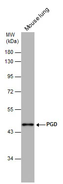
PGD antibody [N1N3] detects PGD protein by western blot analysis. Mouse tissue extracts (50 μg) was separated by 10% SDS-PAGE, and the membrane was blotted with PGD antibody [N1N3] (GTX101703) diluted at 1:500. The HRP-conjugated anti-rabbit IgG antibody (GTX213110-01) was used to detect the primary antibody.
![PGD antibody [N1N3] detects PGD protein at cytoplasm by immunofluorescent analysis. Sample: HeLa cells were fixed in ice-cold MeOH for 5 min. Green: PGD protein stained by PGD antibody [N1N3] (GTX101703) diluted at 1:500. Blue: Hoechst 33342 staining.](https://www.genetex.com/upload/website/prouct_img/normal/GTX101703/GTX101703_39946_IFA_w_23060100_425.webp "PGD antibody [N1N3] detects PGD protein at cytoplasm by immunofluorescent analysis. Sample: HeLa cells were fixed in ice-cold MeOH for 5 min. Green: PGD protein stained by PGD antibody [N1N3] (GTX101703) diluted at 1:500. Blue: Hoechst 33342 staining.")
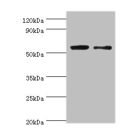
 A:293T B:MOLT4 (GTX27912) C:Raji (GTX27908) 10% SDS PAGE GTX101703 diluted at 1:1000 The HRP-conjugated anti-rabbit IgG antibody (GTX213110-01) was used to detect the primary antibody.")
![PGD antibody [N1N3] detects PGD protein at cytoplasm by immunofluorescent analysis. Sample: HeLa cells were fixed in 4% paraformaldehyde at RT for 15 min. Green: PGD protein stained by PGD antibody [N1N3] (GTX101703) diluted at 1:1000. Red: alpha Tubulin, a cytoskeleton marker, stained by alpha Tubulin antibody [GT114] (GTX628802) diluted at 1:1000. Blue: Hoechst 33342 staining.](https://www.genetex.com/upload/website/prouct_img/normal/GTX101703/GTX101703_39946_20150410_IFA_w_23060100_834.webp "PGD antibody [N1N3] detects PGD protein at cytoplasm by immunofluorescent analysis. Sample: HeLa cells were fixed in 4% paraformaldehyde at RT for 15 min. Green: PGD protein stained by PGD antibody [N1N3] (GTX101703) diluted at 1:1000. Red: alpha Tubulin, a cytoskeleton marker, stained by alpha Tubulin antibody [GT114] (GTX628802) diluted at 1:1000. Blue: Hoechst 33342 staining.")
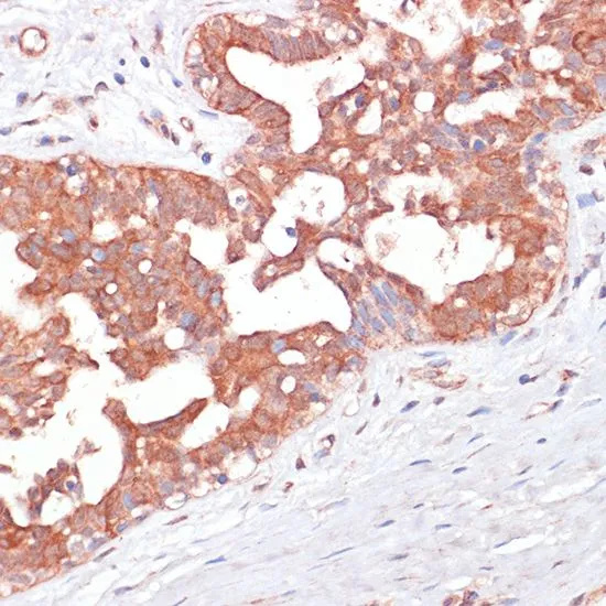
![PGD antibody [N1N3] detects PGD protein at cytoplasm on A549 xenograft by immunohistochemical analysis. Sample: Paraffin-embedded A549 xenograft. PGD antibody [N1N3] (GTX101703) dilution: 1:500.
Antigen Retrieval: Trilogy? (EDTA based, pH 8.0) buffer, 15min](https://www.genetex.com/upload/website/prouct_img/normal/GTX101703/GTX101703_IHC_w_23060100_870.webp "PGD antibody [N1N3] detects PGD protein at cytoplasm on A549 xenograft by immunohistochemical analysis. Sample: Paraffin-embedded A549 xenograft. PGD antibody [N1N3] (GTX101703) dilution: 1:500.
Antigen Retrieval: Trilogy? (EDTA based, pH 8.0) buffer, 15min")
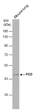
![PGD antibody [N1N3] detects PGD protein by western blot analysis. Mouse tissue extracts (50 μg) was separated by 10% SDS-PAGE, and the membrane was blotted with PGD antibody [N1N3] (GTX101703) diluted at 1:1000. The HRP-conjugated anti-rabbit IgG antibody (GTX213110-01) was used to detect the primary antibody.](https://www.genetex.com/upload/website/prouct_img/normal/GTX101703/GTX101703_39946_20151015_WB_M_heart_w_23060100_278.webp "PGD antibody [N1N3] detects PGD protein by western blot analysis. Mouse tissue extracts (50 μg) was separated by 10% SDS-PAGE, and the membrane was blotted with PGD antibody [N1N3] (GTX101703) diluted at 1:1000. The HRP-conjugated anti-rabbit IgG antibody (GTX213110-01) was used to detect the primary antibody.")

PGD antibody [N1N3] detects PGD protein by western blot analysis. Mouse tissue extracts (50 μg) was separated by 10% SDS-PAGE, and the membrane was blotted with PGD antibody [N1N3] (GTX101703) diluted at 1:500. The HRP-conjugated anti-rabbit IgG antibody (GTX213110-01) was used to detect the primary antibody.
PGD antibody [N1N3]
GTX101703
ApplicationsImmunoFluorescence, Western Blot, ImmunoCytoChemistry, ImmunoHistoChemistry, ImmunoHistoChemistry Paraffin
Product group Antibodies
ReactivityHuman, Mouse
TargetPGD
Overview
- SupplierGeneTex
- Product NamePGD antibody [N1N3]
- Delivery Days Customer9
- Application Supplier NoteWB: 1:500-1:3000. ICC/IF: 1:100-1:1000. IHC-P: 1:100-1:1000. *Optimal dilutions/concentrations should be determined by the researcher.Not tested in other applications.
- ApplicationsImmunoFluorescence, Western Blot, ImmunoCytoChemistry, ImmunoHistoChemistry, ImmunoHistoChemistry Paraffin
- CertificationResearch Use Only
- ClonalityPolyclonal
- Concentration1 mg/ml
- ConjugateUnconjugated
- Gene ID5226
- Target namePGD
- Target descriptionphosphogluconate dehydrogenase
- Target synonyms6PGD, 6-phosphogluconate dehydrogenase, decarboxylating
- HostRabbit
- IsotypeIgG
- Protein IDP52209
- Protein Name6-phosphogluconate dehydrogenase, decarboxylating
- Scientific Description6-phosphogluconate dehydrogenase is the second dehydrogenase in the pentose phosphate shunt. Deficiency of this enzyme is generally asymptomatic, and the inheritance of this disorder is autosomal dominant. Hemolysis results from combined deficiency of 6-phosphogluconate dehydrogenase and 6-phosphogluconolactonase suggesting a synergism of the two enzymopathies. [provided by RefSeq]
- ReactivityHuman, Mouse
- Storage Instruction-20°C or -80°C,2°C to 8°C
- UNSPSC41116161
Datasheet
Related products
Product group Antibodies
PGD AntibodyCSB-PA01155A0RB
ApplicationsWestern Blot, ELISA
ReactivityHuman
TargetPGD
- SizePrice

Product group Antibodies
Anti-PGD Antibody Picoband(r)A01623-3-CARRIER-FREE
ApplicationsFlow Cytometry, ImmunoFluorescence, Western Blot, ELISA, ImmunoCytoChemistry
ReactivityHuman, Mouse, Rat
TargetPGD
- SizePrice
Product group Antibodies
Anti-PGD AntibodyA98459
ApplicationsWestern Blot, ELISA
ReactivityHuman, Mouse, Rat
- SizePrice

Product group Antibodies
Anti-PGD AntibodyHPA031314
ApplicationsWestern Blot, ImmunoHistoChemistry
ReactivityHuman, Mouse
TargetPGD
- SizePrice
Product group Antibodies

PGD AntibodyLS-C349280
ApplicationsWestern Blot, ImmunoHistoChemistry
ReactivityHuman, Mouse, Rat
TargetPGD
- SizePrice
Product group Antibodies
PGD Polyclonal AntibodyCAC13776
ApplicationsWestern Blot, ELISA
TargetPGD
- SizePrice
Product group Antibodies
PGD antibodyGTX101704
ApplicationsImmunoFluorescence, Western Blot, ImmunoCytoChemistry, ImmunoHistoChemistry, ImmunoHistoChemistry Paraffin
ReactivityHuman, Mouse
TargetPGD
- SizePrice
Product group Antibodies
PGD antibodyGTX55744
ApplicationsWestern Blot, ImmunoHistoChemistry, ImmunoHistoChemistry Paraffin
ReactivityHuman, Mouse, Rat
TargetPGD
- SizePrice