

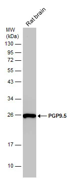
Rat tissue extract (50 μg) was separated by 12% SDS-PAGE, and the membrane was blotted with PGP9.5 antibody (GTX101093) diluted at 1:1000.
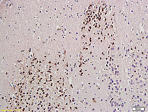
 antibody at 1:100 dilution.
Antigen Retrieval: Trilogy? (EDTA based, pH 8.0) buffer, 15min")
 A: Mouse brain 12% SDS PAGE GTX101093 diluted at 1:1000")
 was separated by 12% SDS-PAGE, and the membrane was blotted with PGP9.5 antibody (GTX101093) diluted at 1:1000. The HRP-conjugated anti-rabbit IgG antibody (GTX213110-01) was used to detect the primary antibody.")
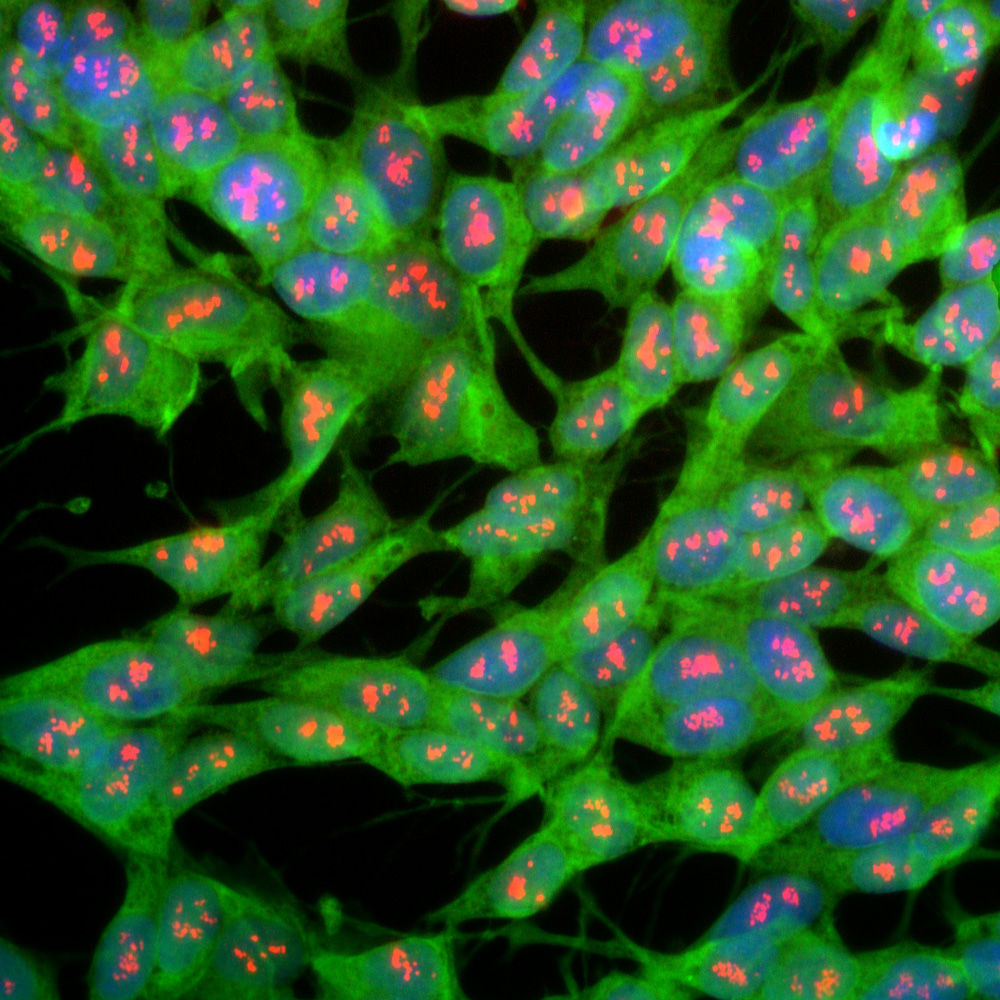
![PGP9.5 antibody detects PGP9.5 protein expression by immunohistochemical analysis. Sample: Frozen sectioned E13.5 Rat brain. Green: PGP9.5 protein stained by PGP9.5 antibody (GTX101093) diluted at 1:250. Red: beta Tubulin 3/ TUJ1, a mature neuron marker, stained by beta Tubulin 3/ TUJ1 antibody [GT11710] (GTX631836) diluted at 1:500. Blue: Fluoroshield with DAPI (GTX30920).](https://www.genetex.com/upload/website/prouct_img/normal/GTX101093/GTX101093_39834_20161012_IHC-Fr_R_w_23060100_140.webp "PGP9.5 antibody detects PGP9.5 protein expression by immunohistochemical analysis. Sample: Frozen sectioned E13.5 Rat brain. Green: PGP9.5 protein stained by PGP9.5 antibody (GTX101093) diluted at 1:250. Red: beta Tubulin 3/ TUJ1, a mature neuron marker, stained by beta Tubulin 3/ TUJ1 antibody [GT11710] (GTX631836) diluted at 1:500. Blue: Fluoroshield with DAPI (GTX30920).")
Rat tissue extract (50 μg) was separated by 12% SDS-PAGE, and the membrane was blotted with PGP9.5 antibody (GTX101093) diluted at 1:1000.
PGP9.5 antibody
GTX101093
ApplicationsWestern Blot, ImmunoHistoChemistry, ImmunoHistoChemistry Frozen, ImmunoHistoChemistry Paraffin
Product group Antibodies
ReactivityHuman, Mouse, Rat
TargetUCHL1
Overview
- SupplierGeneTex
- Product NamePGP9.5 antibody
- Delivery Days Customer9
- Application Supplier NoteWB: 1:500-1:10000. IHC-P: 1:100-1:1000. IHC-Fr: 1:100-1:1000. *Optimal dilutions/concentrations should be determined by the researcher.Not tested in other applications.
- ApplicationsWestern Blot, ImmunoHistoChemistry, ImmunoHistoChemistry Frozen, ImmunoHistoChemistry Paraffin
- CertificationResearch Use Only
- ClonalityPolyclonal
- Concentration0.56 mg/ml
- ConjugateUnconjugated
- Gene ID7345
- Target nameUCHL1
- Target descriptionubiquitin C-terminal hydrolase L1
- Target synonymsHEL-117, HEL-S-53, NDGOA, PARK5, PGP 9.5, PGP9.5, PGP95, SPG79, SPG79A, UCHL-1, Uch-L1, ubiquitin carboxyl-terminal hydrolase isozyme L1, epididymis luminal protein 117, epididymis secretory protein Li 53, neuron cytoplasmic protein 9.5, ubiquitin carboxyl-terminal esterase L1 (ubiquitin thiolesterase), ubiquitin thioesterase L1, ubiquitin thiolesterase
- HostRabbit
- IsotypeIgG
- Protein IDP09936
- Protein NameUbiquitin carboxyl-terminal hydrolase isozyme L1
- Scientific DescriptionUCHL1 is a member of a gene family whose products hydrolyze small C-terminal adducts of ubiquitin to generate the ubiquitin monomer. Expression of UCHL1 is highly specific to neurons and to cells of the diffuse neuroendocrine system and their tumors. It is present in all neurons (Doran et al., 1983 [PubMed 6343558]).[supplied by OMIM]
- ReactivityHuman, Mouse, Rat
- Storage Instruction-20°C or -80°C,2°C to 8°C
- UNSPSC41116161
Datasheet
Related products
Product group Antibodies
Anti-UCHL1 AntibodyA85348
ApplicationsImmunoFluorescence, Western Blot, ImmunoCytoChemistry, ImmunoHistoChemistry
ReactivityBovine, Chicken, Equine, Human, Mouse, Porcine, Rat
- SizePrice
Product group Antibodies
Anti-UCH-L1 Antibody130-10634
ApplicationsELISA
ReactivityHuman
TargetUCHL1
- SizePrice

Product group Antibodies
Anti-UCHL1 AntibodyAMAB91145
ApplicationsWestern Blot, ImmunoCytoChemistry, ImmunoHistoChemistry
ReactivityHuman, Mouse, Rat
TargetUCHL1
- SizePrice
Product group Antibodies
References
PGP9.5 Polyclonal AntibodyBS-3806R
ApplicationsImmunoFluorescence, Western Blot, ELISA, ImmunoCytoChemistry, ImmunoHistoChemistry, ImmunoHistoChemistry Frozen, ImmunoHistoChemistry Paraffin
ReactivityBovine, Equine, Guinea Pig, Human, Mouse, Porcine, Rat
TargetUCHL1
- SizePrice
Product group Antibodies
UCHL1 AntibodyCSB-PA004381
ApplicationsWestern Blot, ELISA, ImmunoHistoChemistry
ReactivityHuman, Mouse, Rat
TargetUCHL1
- SizePrice
Product group Antibodies

Goat anti-UCHL1 (aa 58-68)EB10219
ApplicationsWestern Blot, ELISA, ImmunoHistoChemistry
ReactivityBovine, Canine, Human, Mouse, Porcine, Rat
TargetUCHL1
- SizePrice
Product group Antibodies
ApplicationsFlow Cytometry
TargetUCHL1
- SizePrice
Product group Antibodies
PGP9.5 antibody [ZM160]GTX01799
ApplicationsImmunoHistoChemistry, ImmunoHistoChemistry Paraffin
ReactivityHuman
TargetUCHL1
- SizePrice
![IHC-P analysis of human cerebellum tissue section using GTX02737 PGP9.5 antibody [UCHL1/4556R].](https://www.genetex.com/upload/website/prouct_img/normal/GTX02737/GTX02737_20210319_IHC-P_1_w_23053122_123.webp)
Product group Antibodies
PGP9.5 antibody [UCHL1/4556R]GTX02737
ApplicationsWestern Blot, ImmunoHistoChemistry, ImmunoHistoChemistry Paraffin
ReactivityHuman, Rat
TargetUCHL1
- SizePrice