

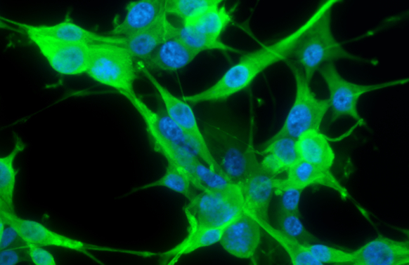
PGP9.5 antibody detects PGP9.5 protein at cytoplasm by immunofluorescent analysis. Sample: U87-MG cells were fixed in 4% paraformaldehyde at RT for 15 min. Green: PGP9.5 stained by PGP9.5 antibody (GTX109637) diluted at 1:200. Blue: Fluoroshield with DAPI (GTX30920).
![PGP9.5 antibody detects PGP9.5 protein at cytoplasm by immunofluorescent analysis. Sample: DIV9 rat E18 primary cortical neurons were fixed in 4% paraformaldehyde at RT for 15 min. Green: PGP9.5 protein stained by PGP9.5 antibody (GTX109637) diluted at 1:500. Red: beta Tubulin 3/ Tuj1, stained by beta Tubulin 3/ Tuj1 antibody [GT11710] (GTX631836) diluted at 1:500. Blue: Fluoroshield with DAPI (GTX30920).](https://www.genetex.com/upload/website/prouct_img/normal/GTX109637/GTX109637_41101_20170503_IFA_R_w_23060500_259.webp "PGP9.5 antibody detects PGP9.5 protein at cytoplasm by immunofluorescent analysis. Sample: DIV9 rat E18 primary cortical neurons were fixed in 4% paraformaldehyde at RT for 15 min. Green: PGP9.5 protein stained by PGP9.5 antibody (GTX109637) diluted at 1:500. Red: beta Tubulin 3/ Tuj1, stained by beta Tubulin 3/ Tuj1 antibody [GT11710] (GTX631836) diluted at 1:500. Blue: Fluoroshield with DAPI (GTX30920).")
![PGP9.5 antibody detects PGP9.5 protein expression by immunohistochemical analysis. Sample: Frozen sectioned E13.5 Rat brain. Green: PGP9.5 protein stained by PGP9.5 antibody (GTX109637) diluted at 1:250. Red: beta Tubulin 3/ TUJ1, a mature neuron marker, stained by beta Tubulin 3/ TUJ1 antibody [GT11710] (GTX631836) diluted at 1:500. Blue: Fluoroshield with DAPI (GTX30920).](https://www.genetex.com/upload/website/prouct_img/normal/GTX109637/GTX109637_41101_20161012_IHC-Fr_R_w_23060500_882.webp "PGP9.5 antibody detects PGP9.5 protein expression by immunohistochemical analysis. Sample: Frozen sectioned E13.5 Rat brain. Green: PGP9.5 protein stained by PGP9.5 antibody (GTX109637) diluted at 1:250. Red: beta Tubulin 3/ TUJ1, a mature neuron marker, stained by beta Tubulin 3/ TUJ1 antibody [GT11710] (GTX631836) diluted at 1:500. Blue: Fluoroshield with DAPI (GTX30920).")
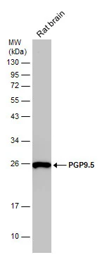
 were separated by 12% SDS-PAGE, and the membrane was blotted with PGP9.5 antibody (GTX109637) diluted at 1:1000. The HRP-conjugated anti-rabbit IgG antibody (GTX213110-01) was used to detect the primary antibody. Corresponding RNA expression data for the same cell lines are based on Human Protein Atlas program.")
![PGP9.5 antibody detects PGP9.5 protein by immunohistochemical analysis. Samples: Paraffin-embedded mouse skin. Green: PGP9.5 protein stained by PGP9.5 antibody (GTX109637) diluted at 1:250. Red: beta Tubulin 3/ Tuj1, a marker, stained by beta Tubulin 3/ Tuj1 antibody [GT1338] (GTX631831) diluted at 1:500. Blue: Fluoroshield with DAPI (GTX30920).
Antigen Retrieval: Citrate buffer, pH 6.0, 15 min](https://www.genetex.com/upload/website/prouct_img/normal/GTX109637/GTX109637_41101_20171127_IHC-P_M_w_23060500_647.webp "PGP9.5 antibody detects PGP9.5 protein by immunohistochemical analysis. Samples: Paraffin-embedded mouse skin. Green: PGP9.5 protein stained by PGP9.5 antibody (GTX109637) diluted at 1:250. Red: beta Tubulin 3/ Tuj1, a marker, stained by beta Tubulin 3/ Tuj1 antibody [GT1338] (GTX631831) diluted at 1:500. Blue: Fluoroshield with DAPI (GTX30920).
Antigen Retrieval: Citrate buffer, pH 6.0, 15 min")
![PGP9.5 antibody detects PGP9.5 protein expression by immunohistochemical analysis. Sample: Frozen-sectioned adult mouse cerebellum. Green: PGP9.5 protein stained by PGP9.5 antibody (GTX109637) diluted at 1:250. Red: beta Tubulin 3/ TUJ1, stained by beta Tubulin 3/ TUJ1 antibody [GT11710] (GTX631836) diluted at 1:500. Blue: Fluoroshield with DAPI (GTX30920).](https://www.genetex.com/upload/website/prouct_img/normal/GTX109637/GTX109637_41101_20170531_IHC-Fr_M_2_w_23060500_314.webp "PGP9.5 antibody detects PGP9.5 protein expression by immunohistochemical analysis. Sample: Frozen-sectioned adult mouse cerebellum. Green: PGP9.5 protein stained by PGP9.5 antibody (GTX109637) diluted at 1:250. Red: beta Tubulin 3/ TUJ1, stained by beta Tubulin 3/ TUJ1 antibody [GT11710] (GTX631836) diluted at 1:500. Blue: Fluoroshield with DAPI (GTX30920).")
![PGP9.5 antibody detects PGP9.5 protein by immunohistochemical analysis. Samples: Paraffin-embedded rat colon. Green: PGP9.5 protein stained by PGP9.5 antibody (GTX109637) diluted at 1:250. Red: beta Tubulin 3/ Tuj1, a marker, stained by beta Tubulin 3/ Tuj1 antibody [GT1338] (GTX631831) diluted at 1:500. Blue: Fluoroshield with DAPI (GTX30920).
Antigen Retrieval: Citrate buffer, pH 6.0, 15 min](https://www.genetex.com/upload/website/prouct_img/normal/GTX109637/GTX109637_41101_20171127_IHC-P_R_w_23060500_414.webp "PGP9.5 antibody detects PGP9.5 protein by immunohistochemical analysis. Samples: Paraffin-embedded rat colon. Green: PGP9.5 protein stained by PGP9.5 antibody (GTX109637) diluted at 1:250. Red: beta Tubulin 3/ Tuj1, a marker, stained by beta Tubulin 3/ Tuj1 antibody [GT1338] (GTX631831) diluted at 1:500. Blue: Fluoroshield with DAPI (GTX30920).
Antigen Retrieval: Citrate buffer, pH 6.0, 15 min")
 was separated by 12% SDS-PAGE, and the membrane was blotted with PGP9.5 antibody (GTX109637) diluted at 1:10000.")
 and transfected (+) 293T whole cell extracts (30 μg) were separated by 12% SDS-PAGE, and the membrane was blotted with PGP9.5 antibody (GTX109637) diluted at 1:3000. The HRP-conjugated anti-rabbit IgG antibody (GTX213110-01) was used to detect the primary antibody.")
 dilution: 1:10000 The HRP-conjugated anti-rabbit IgG antibody (GTX213110-01) was used to detect the primary antibody.")
PGP9.5 antibody detects PGP9.5 protein at cytoplasm by immunofluorescent analysis. Sample: U87-MG cells were fixed in 4% paraformaldehyde at RT for 15 min. Green: PGP9.5 stained by PGP9.5 antibody (GTX109637) diluted at 1:200. Blue: Fluoroshield with DAPI (GTX30920).
PGP9.5 antibody
GTX109637
ApplicationsImmunoFluorescence, Western Blot, ImmunoCytoChemistry, ImmunoHistoChemistry, ImmunoHistoChemistry Frozen, ImmunoHistoChemistry Paraffin
Product group Antibodies
ReactivityHuman, Mouse, Rat
TargetUCHL1
Overview
- SupplierGeneTex
- Product NamePGP9.5 antibody
- Delivery Days Customer9
- Application Supplier NoteWB: 1:1000-1:20000. ICC/IF: 1:100-1:1000. IHC-P: 1:100-1:1000. IHC-Fr: 1:100-1:1000. *Optimal dilutions/concentrations should be determined by the researcher.Not tested in other applications.
- ApplicationsImmunoFluorescence, Western Blot, ImmunoCytoChemistry, ImmunoHistoChemistry, ImmunoHistoChemistry Frozen, ImmunoHistoChemistry Paraffin
- CertificationResearch Use Only
- ClonalityPolyclonal
- Concentration1.79 mg/ml
- ConjugateUnconjugated
- Gene ID7345
- Target nameUCHL1
- Target descriptionubiquitin C-terminal hydrolase L1
- Target synonymsHEL-117, HEL-S-53, NDGOA, PARK5, PGP 9.5, PGP9.5, PGP95, SPG79, SPG79A, UCHL-1, Uch-L1, ubiquitin carboxyl-terminal hydrolase isozyme L1, epididymis luminal protein 117, epididymis secretory protein Li 53, neuron cytoplasmic protein 9.5, ubiquitin carboxyl-terminal esterase L1 (ubiquitin thiolesterase), ubiquitin thioesterase L1, ubiquitin thiolesterase
- HostRabbit
- IsotypeIgG
- Protein IDP09936
- Protein NameUbiquitin carboxyl-terminal hydrolase isozyme L1
- Scientific DescriptionUCHL1 is a member of a gene family whose products hydrolyze small C-terminal adducts of ubiquitin to generate the ubiquitin monomer. Expression of UCHL1 is highly specific to neurons and to cells of the diffuse neuroendocrine system and their tumors. It is present in all neurons (Doran et al., 1983 [PubMed 6343558]).[supplied by OMIM]
- ReactivityHuman, Mouse, Rat
- Storage Instruction-20°C or -80°C,2°C to 8°C
- UNSPSC12352203
References
- Clayton DR, Ruiz WG, Dalghi MG, et al. Studies of ultrastructure, gene expression, and marker analysis reveal that mouse bladder PDGFRA(+) interstitial cells are fibroblasts. Am J Physiol Renal Physiol. 2022,323(3):F299-F321. doi: 10.1152/ajprenal.00135.2022Read this paper
- Chang WY, Yang YT, She MP, et al. 5-HT(7) receptor-dependent intestinal neurite outgrowth contributes to visceral hypersensitivity in irritable bowel syndrome. Lab Invest. 2022,102(9):1023-1037. doi: 10.1038/s41374-022-00800-zRead this paper
- Fede C, Petrelli L, Guidolin D, et al. Evidence of a new hidden neural network into deep fasciae. Sci Rep. 2021,11(1):12623. doi: 10.1038/s41598-021-92194-zRead this paper
- Koshimizu H, Ohkawara B, Nakashima H, et al. Zonisamide ameliorates neuropathic pain partly by suppressing microglial activation in the spinal cord in a mouse model. Life Sci. 2020,263:118577. doi: 10.1016/j.lfs.2020.118577Read this paper
- Xu YM, Tan HW, Zheng W, et al. Cadmium telluride quantum dot-exposed human bronchial epithelial cells: a further study of the cellular response by proteomics. Toxicol Res (Camb). 2019,8(6):994-1001. doi: 10.1039/c9tx00126cRead this paper
- Hong J, Lisco AM, Rudebush TL, et al. Identification of Cardiac Expression Pattern of Transient Receptor Potential Vanilloid Type 1 (TRPV1) Receptor using a Transgenic Reporter Mouse Model. Neurosci Lett. 2020,737:135320. doi: 10.1016/j.neulet.2020.135320Read this paper
- Canta A, Chiorazzi A, Pozzi E, et al. Calmangafodipir Reduces Sensory Alterations and Prevents Intraepidermal Nerve Fibers Loss in a Mouse Model of Oxaliplatin Induced Peripheral Neurotoxicity. Antioxidants (Basel). 2020,9(7). doi: 10.3390/antiox9070594Read this paper
- Dragunas G, Woest ME, Nijboer S, et al. Cholinergic neuroplasticity in asthma driven by TrkB signaling. FASEB J. 2020,34(6):7703-7717. doi: 10.1096/fj.202000170RRead this paper
- Pagella P, Catón J, Meisel CT, et al. Ameloblastomas Exhibit Stem Cell Potential, Possess Neurotrophic Properties, and Establish Connections with Trigeminal Neurons. Cells. 2020,9(3). doi: 10.3390/cells9030644Read this paper
- Alberti P, Canta A, Chiorazzi A, et al. Topiramate prevents oxaliplatin-related axonal hyperexcitability and oxaliplatin induced peripheral neurotoxicity. Neuropharmacology. 2020,164:107905. doi: 10.1016/j.neuropharm.2019.107905Read this paper
Datasheet
Related products
Product group Antibodies
Anti-UCH-L1 Antibody130-10634
ApplicationsELISA
ReactivityHuman
TargetUCHL1
- SizePrice
![IHC-P analysis of human cerebellum tissue section using GTX02737 PGP9.5 antibody [UCHL1/4556R].](https://www.genetex.com/upload/website/prouct_img/normal/GTX02737/GTX02737_20210319_IHC-P_1_w_23053122_123.webp)
Product group Antibodies
PGP9.5 antibody [UCHL1/4556R]GTX02737
ApplicationsWestern Blot, ImmunoHistoChemistry, ImmunoHistoChemistry Paraffin
ReactivityHuman, Rat
TargetUCHL1
- SizePrice
![IHC-P analysis of human testis tissue using GTX04438 PGP9.5 antibody [MSVA-905R] HistoMAX?. A strong PGP9.5 immunostaining is seen in spermatogonia. PGP9.5 staining decreases sharply in spermatocytes where it is only faint. A moderate PGP9.5 staining is seen in Leydig cells while Sertoli cells remain PGP9.5 negative.](https://www.genetex.com/upload/website/prouct_img/normal/GTX04438/GTX04438_20230728_IHC-P_97_23072722_457.webp)
Product group Antibodies
ApplicationsImmunoHistoChemistry, ImmunoHistoChemistry Paraffin
ReactivityHuman
TargetUCHL1
- SizePrice
Product group Antibodies
PGP9.5 antibodyGTX101093
ApplicationsWestern Blot, ImmunoHistoChemistry, ImmunoHistoChemistry Frozen, ImmunoHistoChemistry Paraffin
ReactivityHuman, Mouse, Rat
TargetUCHL1
- SizePrice
Product group Antibodies
PGP9.5 antibodyGTX10404
ApplicationsWestern Blot, ImmunoHistoChemistry, ImmunoHistoChemistry Frozen
ReactivityHuman, Mouse, Porcine, Rat
TargetUCHL1
- SizePrice
![PGP9.5 antibody detects PGP9.5 protein at cytoplasm by immunofluorescent analysis. Sample: DIV9 rat E18 primary cortical neurons were fixed in 4% paraformaldehyde at RT for 15 min. Green: PGP9.5 protein stained by PGP9.5 antibody (GTX109646) diluted at 1:500. Red: beta Tubulin 3/ Tuj1, stained by beta Tubulin 3/ Tuj1 antibody [GT11710] (GTX631836) diluted at 1:500. Blue: Fluoroshield with DAPI (GTX30920).](https://www.genetex.com/upload/website/prouct_img/normal/GTX109646/GTX109646_39959_20170503_IFA_R_w_23060500_628.webp)
Product group Antibodies
PGP9.5 antibodyGTX109646
ApplicationsImmunoFluorescence, Western Blot, ImmunoCytoChemistry, ImmunoHistoChemistry, ImmunoHistoChemistry Frozen, ImmunoHistoChemistry Paraffin
ReactivityHuman, Mouse, Rat
TargetUCHL1
- SizePrice
Product group Antibodies
PGP9.5 antibody [3D9]GTX57556
ApplicationsImmunoFluorescence, Western Blot, ImmunoCytoChemistry, ImmunoHistoChemistry, ImmunoHistoChemistry Paraffin
ReactivityHuman, Mouse
TargetUCHL1
- SizePrice
![PGP9.5 antibody [GT448] detects PGP9.5 protein by immunofluorescent analysis. Sample: DIV9 rat E18 primary hippocampal neuron cells were fixed in 4% paraformaldehyde at RT for 15 min. Green: PGP9.5 stained by PGP9.5 antibody [GT448] (GTX634797) diluted at 1:500. Red: NeuN, stained by NeuN antibody (GTX132974) diluted at 1:1000. Blue: Fluoroshield with DAPI (GTX30920).](https://www.genetex.com/upload/website/prouct_img/normal/GTX634797/GTX634797_43339_20190306_ICC_IF_R_w_23061202_510.webp)
Product group Antibodies
PGP9.5 antibody [GT448]GTX634797
ApplicationsImmunoFluorescence, Western Blot, ImmunoCytoChemistry, ImmunoHistoChemistry, ImmunoHistoChemistry Paraffin
ReactivityHuman, Mouse, Rat
TargetUCHL1
- SizePrice
Product group Antibodies
Anti-UCHL1Y058669
ApplicationsWestern Blot, ELISA, ImmunoHistoChemistry
ReactivityHuman
- SizePrice