

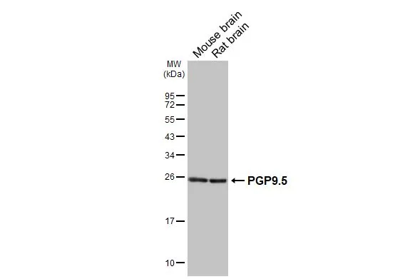
Various tissue extracts (50 μg) were separated by 12% SDS-PAGE, and the membrane was blotted with PGP9.5 antibody [HL3391] (GTX641211) diluted at 1:1000. The HRP-conjugated anti-rabbit IgG antibody (GTX213110-01) was used to detect the primary antibody.
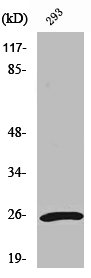
![Various whole cell extracts (30 μg) were separated by 12% SDS-PAGE, and the membrane was blotted with PGP9.5 antibody [HL3391] (GTX641211) diluted at 1:1000. The HRP-conjugated anti-rabbit IgG antibody (GTX213110-01) was used to detect the primary antibody. Corresponding RNA expression data for the same cell lines are based on Human Protein Atlas program.](https://www.genetex.com/upload/website/prouct_img/normal/GTX641211/GTX641211_T-45572_20241025_WB_TPM_watermark_24103022_462.webp "Various whole cell extracts (30 μg) were separated by 12% SDS-PAGE, and the membrane was blotted with PGP9.5 antibody [HL3391] (GTX641211) diluted at 1:1000. The HRP-conjugated anti-rabbit IgG antibody (GTX213110-01) was used to detect the primary antibody. Corresponding RNA expression data for the same cell lines are based on Human Protein Atlas program.")
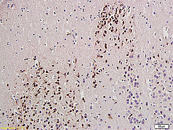
![PGP9.5 antibody [HL3391] detects PGP9.5 protein by immunohistochemical analysis. Sample: Paraffin-embedded human tissues. PGP9.5 stained by PGP9.5 antibody [HL3391] (GTX641211) diluted at 1:1000. Antigen Retrieval: Citrate buffer, pH 6.0, 15 min](https://www.genetex.com/upload/website/prouct_img/normal/GTX641211/GTX641211_45628_20250509_IHC-P_Multiple_RPKM_25051420_239.webp "PGP9.5 antibody [HL3391] detects PGP9.5 protein by immunohistochemical analysis. Sample: Paraffin-embedded human tissues. PGP9.5 stained by PGP9.5 antibody [HL3391] (GTX641211) diluted at 1:1000. Antigen Retrieval: Citrate buffer, pH 6.0, 15 min")
![PGP9.5 antibody [HL3391] detects PGP9.5 protein by immunohistochemical analysis. Sample: Paraffin-embedded human pancreas. PGP9.5 stained by PGP9.5 antibody [HL3391] (GTX641211) diluted at 1:1000. Antigen Retrieval: Citrate buffer, pH 6.0, 15 min](https://www.genetex.com/upload/website/prouct_img/normal/GTX641211/GTX641211_45628_20250509_IHC-P_25051420_372.webp "PGP9.5 antibody [HL3391] detects PGP9.5 protein by immunohistochemical analysis. Sample: Paraffin-embedded human pancreas. PGP9.5 stained by PGP9.5 antibody [HL3391] (GTX641211) diluted at 1:1000. Antigen Retrieval: Citrate buffer, pH 6.0, 15 min")
![PGP9.5 antibody [HL3391] detects PGP9.5 protein by immunohistochemical analysis. Sample: Paraffin-embedded mouse pancreas. PGP9.5 stained by PGP9.5 antibody [HL3391] (GTX641211) diluted at 1:1000. Antigen Retrieval: Citrate buffer, pH 6.0, 15 min](https://www.genetex.com/upload/website/prouct_img/normal/GTX641211/GTX641211_45628_20250509_IHC-P_M_1_25051420_937.webp "PGP9.5 antibody [HL3391] detects PGP9.5 protein by immunohistochemical analysis. Sample: Paraffin-embedded mouse pancreas. PGP9.5 stained by PGP9.5 antibody [HL3391] (GTX641211) diluted at 1:1000. Antigen Retrieval: Citrate buffer, pH 6.0, 15 min")
![PGP9.5 antibody [HL3391] detects PGP9.5 protein by immunohistochemical analysis. Sample: Paraffin-embedded mouse tissues. PGP9.5 stained by PGP9.5 antibody [HL3391] (GTX641211) diluted at 1:1000. Antigen Retrieval: Citrate buffer, pH 6.0, 15 min](https://www.genetex.com/upload/website/prouct_img/normal/GTX641211/GTX641211_45628_20250509_IHC-P_M_multiple_KPKM_25051420_145.webp "PGP9.5 antibody [HL3391] detects PGP9.5 protein by immunohistochemical analysis. Sample: Paraffin-embedded mouse tissues. PGP9.5 stained by PGP9.5 antibody [HL3391] (GTX641211) diluted at 1:1000. Antigen Retrieval: Citrate buffer, pH 6.0, 15 min")
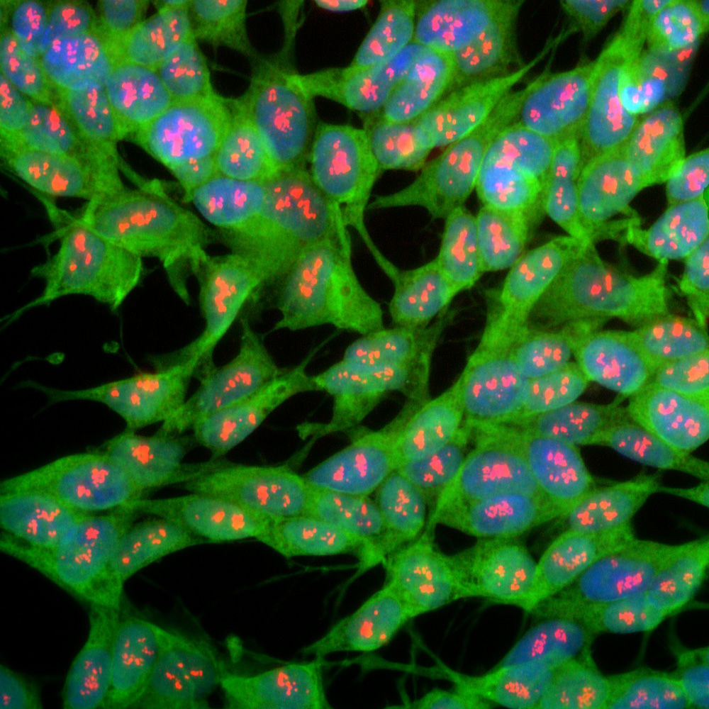
![PGP9.5 antibody [HL3391] detects PGP9.5 protein by immunohistochemical analysis. Sample: Paraffin-embedded mouse eye. Green: PGP9.5 stained by PGP9.5 antibody [HL3391] (GTX641211) diluted at 1:1000. Red: beta Tubulin 3/ Tuj1 antibody [GT11710] (GTX631836) diluted at 1:500. Blue: Fluoroshield with DAPI (GTX30920). Antigen Retrieval: Citrate buffer, pH 6.0, 15 min](https://www.genetex.com/upload/website/prouct_img/normal/GTX641211/GTX641211_45628_20250509_IHC-P_M_2_25051420_402.webp "PGP9.5 antibody [HL3391] detects PGP9.5 protein by immunohistochemical analysis. Sample: Paraffin-embedded mouse eye. Green: PGP9.5 stained by PGP9.5 antibody [HL3391] (GTX641211) diluted at 1:1000. Red: beta Tubulin 3/ Tuj1 antibody [GT11710] (GTX631836) diluted at 1:500. Blue: Fluoroshield with DAPI (GTX30920). Antigen Retrieval: Citrate buffer, pH 6.0, 15 min")
![PGP9.5 antibody [HL3391] detects PGP9.5 protein by immunohistochemical analysis. Sample: Frozen-sectioned mouse colon. Green: PGP9.5 stained by PGP9.5 antibody [HL3391] (GTX641211) diluted at 1:250. Red: beta Tubulin 3/ Tuj1 antibody [GT11710] (GTX631836) diluted at 1:500. Blue: Hoechst 33342 staining.](https://www.genetex.com/upload/website/prouct_img/normal/GTX641211/GTX641211_45628_20250516_IHC-Fr_M_25061003_260.webp "PGP9.5 antibody [HL3391] detects PGP9.5 protein by immunohistochemical analysis. Sample: Frozen-sectioned mouse colon. Green: PGP9.5 stained by PGP9.5 antibody [HL3391] (GTX641211) diluted at 1:250. Red: beta Tubulin 3/ Tuj1 antibody [GT11710] (GTX631836) diluted at 1:500. Blue: Hoechst 33342 staining.")
Various tissue extracts (50 μg) were separated by 12% SDS-PAGE, and the membrane was blotted with PGP9.5 antibody [HL3391] (GTX641211) diluted at 1:1000. The HRP-conjugated anti-rabbit IgG antibody (GTX213110-01) was used to detect the primary antibody.
PGP9.5 antibody [HL3391]
GTX641211
ApplicationsWestern Blot, ImmunoHistoChemistry, ImmunoHistoChemistry Frozen, ImmunoHistoChemistry Paraffin
Product group Antibodies
ReactivityHuman, Mouse, Rat
TargetUCHL1
Overview
- SupplierGeneTex
- Product NamePGP9.5 antibody [HL3391]
- Delivery Days Customer7
- ApplicationsWestern Blot, ImmunoHistoChemistry, ImmunoHistoChemistry Frozen, ImmunoHistoChemistry Paraffin
- CertificationResearch Use Only
- ClonalityMonoclonal
- Clone IDHL3391
- Concentration1 mg/ml
- ConjugateUnconjugated
- Gene ID7345
- Target nameUCHL1
- Target descriptionubiquitin C-terminal hydrolase L1
- Target synonymsHEL-117, HEL-S-53, NDGOA, PARK5, PGP 9.5, PGP9.5, PGP95, SPG79, SPG79A, UCHL-1, Uch-L1, ubiquitin carboxyl-terminal hydrolase isozyme L1, epididymis luminal protein 117, epididymis secretory protein Li 53, neuron cytoplasmic protein 9.5, ubiquitin carboxyl-terminal esterase L1 (ubiquitin thiolesterase), ubiquitin thioesterase L1, ubiquitin thiolesterase
- HostRabbit
- IsotypeIgG
- Protein IDP09936
- Protein NameUbiquitin carboxyl-terminal hydrolase isozyme L1
- Scientific DescriptionThe protein encoded by this gene belongs to the peptidase C12 family. This enzyme is a thiol protease that hydrolyzes a peptide bond at the C-terminal glycine of ubiquitin. This gene is specifically expressed in the neurons and in cells of the diffuse neuroendocrine system. Mutations in this gene may be associated with Parkinson disease.[provided by RefSeq, Sep 2009]
- ReactivityHuman, Mouse, Rat
- Storage Instruction-20°C or -80°C,2°C to 8°C
- UNSPSC41116161
Datasheet
Related products
Product group Antibodies
UCHL1 AntibodyCSB-PA004381
ApplicationsWestern Blot, ELISA, ImmunoHistoChemistry
ReactivityHuman, Mouse, Rat
TargetUCHL1
- SizePrice
Product group Antibodies
Anti-UCHL1 AntibodyA85348
ApplicationsImmunoFluorescence, Western Blot, ImmunoCytoChemistry, ImmunoHistoChemistry
ReactivityBovine, Chicken, Equine, Human, Mouse, Porcine, Rat
- SizePrice

Product group Antibodies
Anti-UCHL1 AntibodyAMAB91145
ApplicationsWestern Blot, ImmunoCytoChemistry, ImmunoHistoChemistry
ReactivityHuman, Mouse, Rat
TargetUCHL1
- SizePrice
Product group Antibodies

UCHL1 / PGP9.5 AntibodyLS-C835160
ApplicationsImmunoHistoChemistry
ReactivityHuman
TargetUCHL1
- SizePrice
Product group Antibodies

Goat anti-UCHL1 (aa 58-68)EB10219
ApplicationsWestern Blot, ELISA, ImmunoHistoChemistry
ReactivityBovine, Canine, Human, Mouse, Porcine, Rat
TargetUCHL1
- SizePrice
Product group Antibodies
ApplicationsFlow Cytometry
TargetUCHL1
- SizePrice

Product group Antibodies
Anti-PGP9.5/UCHL1 Antibody Picoband(r)PB9840-CARRIER-FREE
ApplicationsFlow Cytometry, ImmunoFluorescence, Western Blot, ImmunoCytoChemistry, ImmunoHistoChemistry
ReactivityHamster, Human, Mouse, Rat
TargetUCHL1
- SizePrice
Product group Antibodies
References
PGP9.5 Polyclonal AntibodyBS-3806R
ApplicationsImmunoFluorescence, Western Blot, ELISA, ImmunoCytoChemistry, ImmunoHistoChemistry, ImmunoHistoChemistry Frozen, ImmunoHistoChemistry Paraffin
ReactivityBovine, Equine, Guinea Pig, Human, Mouse, Porcine, Rat
TargetUCHL1
- SizePrice
Product group Antibodies
PGP9.5 antibody [13C4]GTX72095
ApplicationsImmunoHistoChemistry, ImmunoHistoChemistry Paraffin
ReactivityHuman, Mouse, Rat
TargetUCHL1
- SizePrice