

Phospho-KDR/FLT4 (Y1054/Y1063) Antibody
CSB-PA009643
ApplicationsELISA, ImmunoHistoChemistry
Product group Antibodies
ReactivityHuman, Mouse, Rat
TargetFLT4
Overview
- SupplierCusabio
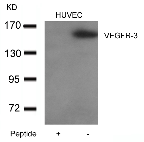
- Product NamePhospho-KDR/FLT4 (Y1054/Y1063) Antibody
- Delivery Days Customer20
- ApplicationsELISA, ImmunoHistoChemistry
- CertificationResearch Use Only
- ClonalityPolyclonal
- ConjugateUnconjugated
- Gene ID2324
- Target nameFLT4
- Target descriptionfms related receptor tyrosine kinase 4
- Target synonymsCHTD7, FLT-4, FLT41, LMPH1A, LMPHM1, PCL, VEGFR-3, VEGFR3, vascular endothelial growth factor receptor 3, Feline McDonough Sarcoma (FMS)-like tyrosine kinase 4, VEGF receptor-3, fms related tyrosine kinase 4, fms-like tyrosine kinase 4, primary congenital lymphedema, tyrosine-protein kinase receptor FLT4
- HostRabbit
- IsotypeIgG
- Protein IDP35916
- Protein NameVascular endothelial growth factor receptor 3
- ReactivityHuman, Mouse, Rat
- Storage Instruction-20°C or -80°C
- UNSPSC41116161
Related products

Product group Antibodies
Anti-VEGF Receptor 3/FLT4 Antibody Picoband(r)A01276-2-CARRIER-FREE
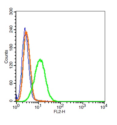
ApplicationsFlow Cytometry, ImmunoFluorescence, Western Blot, ImmunoCytoChemistry, ImmunoHistoChemistry, ImmunoHistoChemistry Frozen
ReactivityHuman, Mouse, Rat
TargetFLT4
- SizePrice
Product group Antibodies

Anti-VEGFR-3 [ABDD073]AB04028-10.0
ApplicationsFlow Cytometry, ELISA, ImmunoHistoChemistry
ReactivityHuman
TargetFLT4
- SizePrice
Product group Antibodies
Anti-VEGFR3 AntibodyA34796
ApplicationsImmunoFluorescence, Western Blot, ImmunoHistoChemistry
ReactivityHuman
- SizePrice
Product group Antibodies

FLT4 / VEGFR3 AntibodyLS-C832540
ApplicationsImmunoFluorescence, ELISA
ReactivityHuman
TargetFLT4
- SizePrice

Product group Antibodies
Anti-FLT4 AntibodyHPA046519
ApplicationsImmunoCytoChemistry
ReactivityHuman
TargetFLT4
- SizePrice
Product group Antibodies
ApplicationsImmunoPrecipitation, Western Blot, ImmunoCytoChemistry, ImmunoHistoChemistry
TargetFLT4
- SizePrice
Product group Antibodies
References
VEGFR3 Polyclonal AntibodyBS-2202R
ApplicationsFlow Cytometry, ImmunoFluorescence, ELISA, ImmunoCytoChemistry, ImmunoHistoChemistry, ImmunoHistoChemistry Frozen, ImmunoHistoChemistry Paraffin
ReactivityCanine, Equine, Human, Mouse, Porcine, Rabbit, Rat
TargetFLT4
- SizePrice
Product group Antibodies
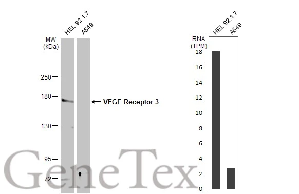
VEGF Receptor 3 antibodyGTX100807
ApplicationsWestern Blot
ReactivityHuman, Mouse
TargetFLT4
- SizePrice
Product group Antibodies
Anti-Mouse/Rat FLT4 Antibody144-12332
ApplicationsWestern Blot
ReactivityHuman, Mouse, Rat
TargetFLT4
- SizePrice