

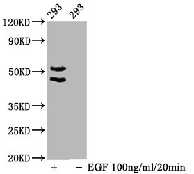
Western Blot Positive WB detected in 293 whole cell lysate(treated with EGF or not) All lanes Phospho-MAPK8/MAPK9/MAPK10 antibody at 1.65microg/ml Secondary Goat polyclonal to rabbit IgG at 1/50000 dilution Predicted band size: 46,54 KDa Observed band size: 46,54 KDa
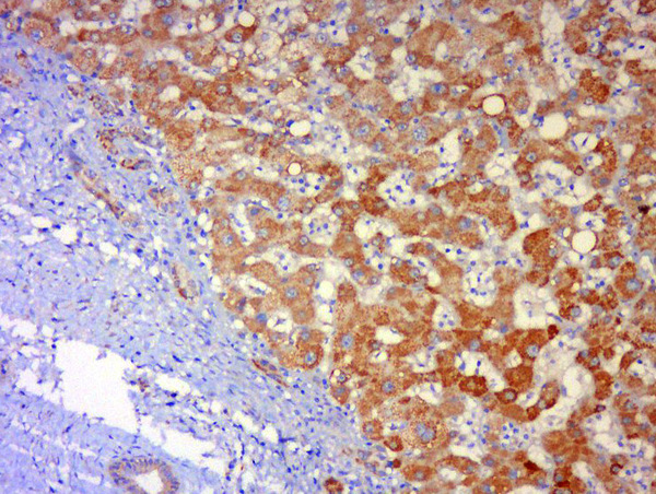
 performed on a Leica BondTM system. The cells were fixed in 4% formaldehyde, permeabilized using 0.2% Triton X-100 and blocked with 10% normal goat serum 30min at RT. Then primary antibody (1% BSA) was incubated at 4°C overnight. The primary is detected by a biotinylated secondary antibody and visualized using an HRP conjugated SP system.")
Western Blot Positive WB detected in 293 whole cell lysate(treated with EGF or not) All lanes Phospho-MAPK8/MAPK9/MAPK10 antibody at 1.65microg/ml Secondary Goat polyclonal to rabbit IgG at 1/50000 dilution Predicted band size: 46,54 KDa Observed band size: 46,54 KDa
Phospho-MAPK8/MAPK9/MAPK10 (T183/T183/T221) Recombinant Monoclonal Antibody
CSB-RA013466A183PHHU
ApplicationsWestern Blot, ELISA, ImmunoHistoChemistry
Product group Antibodies
ReactivityHuman
TargetMAPK8
Overview
- SupplierCusabio
- Product NamePhospho-MAPK8/MAPK9/MAPK10 (T183/T183/T221) Recombinant Monoclonal Antibody
- Delivery Days Customer20
- ApplicationsWestern Blot, ELISA, ImmunoHistoChemistry
- CertificationResearch Use Only
- ClonalityMonoclonal
- Clone ID1A9
- ConjugateUnconjugated
- Gene ID5599
- Target nameMAPK8
- Target descriptionmitogen-activated protein kinase 8
- Target synonymsJNK, JNK-46, JNK1, JNK1A2, JNK21B1/2, PRKM8, SAPK1, SAPK1c, mitogen-activated protein kinase 8, JUN N-terminal kinase, MAP kinase 8, c-Jun N-terminal kinase 1, stress-activated protein kinase 1, stress-activated protein kinase 1c
- IsotypeIgG
- Protein IDP45983
- Protein NameMitogen-activated protein kinase 8
- Scientific DescriptionSerine/threonine-protein kinase involved in various processes such as cell proliferation, differentiation, migration, transformation and programmed cell death. Extracellular stimuli such as proinflammatory cytokines or physical stress stimulate the stress-activated protein kinase/c-Jun N-terminal kinase (SAP/JNK) signaling pathway. In this cascade, two dual specificity kinases MAP2K4/MKK4 and MAP2K7/MKK7 phosphorylate and activate MAPK8/JNK1. In turn, MAPK8/JNK1 phosphorylates a number of transcription factors, primarily components of AP-1 such as JUN, JDP2 and ATF2 and thus regulates AP-1 transcriptional activity. Phosphorylates the replication licensing factor CDT1, inhibiting the interaction between CDT1 and the histone H4 acetylase HBO1 to replication origins. Loss of this interaction abrogates the acetylation required for replication initiation. Promotes stressed cell apoptosis by phosphorylating key regulatory factors including p53/TP53 and Yes-associates protein YAP1. In T-cells, MAPK8 and MAPK9 are required for polarized differentiation of T-helper cells into Th1 cells. Contributes to the survival of erythroid cells by phosphorylating the antagonist of cell death BAD upon EPO stimulation. Mediates starvation-induced BCL2 phosphorylation, BCL2 dissociation from BECN1, and thus activation of autophagy. Phosphorylates STMN2 and hence regulates microtubule dynamics, controlling neurite elongation in cortical neurons. In the developing brain, through its cytoplasmic activity on STMN2, negatively regulates the rate of exit from multipolar stage and of radial migration from the ventricular zone. Phosphorylates several other substrates including heat shock factor protein 4 (HSF4), the deacetylase SIRT1, ELK1, or the E3 ligase ITCH. Phosphorylates the CLOCK-ARNTL/BMAL1 heterodimer and plays a role in the regulation of the circadian clock (PubMed:22441692). Phosphorylates the heat shock transcription factor HSF1, suppressing HSF1-induced transcriptional activity (PubMed:10747973).
- ReactivityHuman
- Storage Instruction-20°C or -80°C
- UNSPSC41116161
Related products
Product group Antibodies
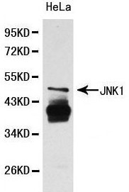
Anti-JNK1 AntibodyA29715
ApplicationsWestern Blot
ReactivityHuman, Mouse, Rat
- SizePrice
Product group Antibodies
Anti-MAPK8 Antibody144-00288
ApplicationsImmunoFluorescence, Western Blot, ImmunoHistoChemistry
ReactivityHuman, Mouse
TargetMAPK8
- SizePrice
Product group Antibodies
References
JNK1 + JNK3 Polyclonal AntibodyBS-0501R
ApplicationsFlow Cytometry, ImmunoFluorescence, Western Blot, ELISA, ImmunoCytoChemistry, ImmunoHistoChemistry, ImmunoHistoChemistry Frozen, ImmunoHistoChemistry Paraffin
ReactivityBovine, Canine, Chicken, Human, Mouse, Porcine, Rabbit, Rat
TargetMAPK8
- SizePrice
Product group Antibodies
MAPK8/MAPK9/MAPK10 AntibodyCSB-PA003084
ApplicationsImmunoFluorescence, Western Blot, ELISA, ImmunoHistoChemistry
ReactivityHuman, Mouse, Rat
TargetMAPK8
- SizePrice
Product group Antibodies
MAPK8 Polyclonal AntibodyCAC14610
ApplicationsImmunoFluorescence, Western Blot, ELISA, ImmunoHistoChemistry
TargetMAPK8
- SizePrice

Product group Antibodies
References
ApplicationsImmunoFluorescence, ImmunoPrecipitation, Western Blot, ImmunoCytoChemistry
ReactivityHuman, Mouse, Rat
TargetMAPK8
- SizePrice
Product group Antibodies
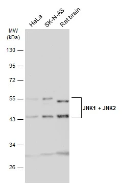
JNK1 + JNK2 antibodyGTX133806
ApplicationsWestern Blot, ImmunoHistoChemistry, ImmunoHistoChemistry Paraffin
ReactivityHuman, Rat
TargetMAPK8
- SizePrice
Product group Antibodies

ApplicationsELISA
ReactivityHuman
TargetMAPK8
- SizePrice