

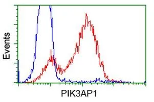
FACS analysis of HEK293T cells transfected with either PIK3AP1 plasmid(Red) or empty vector control plasmid(Blue) using GTX83893 PIK3AP1 antibody [7A11].
![IHC-P analysis of human lymph node tissue using GTX83893 PIK3AP1 antibody [7A11]. Antigen retrieval : Heat-induced epitope retrieval by 10mM citrate buffer, pH6.0, 100oC for 10min.](https://www.genetex.com/upload/website/prouct_img/normal/GTX83893/GTX83893_1992_IHC-P_w_23061420_845.webp "IHC-P analysis of human lymph node tissue using GTX83893 PIK3AP1 antibody [7A11]. Antigen retrieval : Heat-induced epitope retrieval by 10mM citrate buffer, pH6.0, 100oC for 10min.")
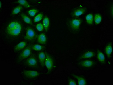
![ICC/IF analysis of COS7 cells transiently transfected with PIK3AP1 plasmid using GTX83893 PIK3AP1 antibody [7A11].](https://www.genetex.com/upload/website/prouct_img/normal/GTX83893/GTX83893_866_ICCIF_w_23061420_900.webp "ICC/IF analysis of COS7 cells transiently transfected with PIK3AP1 plasmid using GTX83893 PIK3AP1 antibody [7A11].")
![WB analysis of HEK293T cells transfected with PIK3AP1 plasmid (Right) or empty vector (Left) for 48 hrs using GTX83893 PIK3AP1 antibody [7A11]. Loading : 5 ug per lane](https://www.genetex.com/upload/website/prouct_img/normal/GTX83893/GTX83893_3975_WB_w_23061420_183.webp "WB analysis of HEK293T cells transfected with PIK3AP1 plasmid (Right) or empty vector (Left) for 48 hrs using GTX83893 PIK3AP1 antibody [7A11]. Loading : 5 ug per lane")
![IHC-P analysis of human lymphoma tissue using GTX83893 PIK3AP1 antibody [7A11]. Antigen retrieval : Heat-induced epitope retrieval by 10mM citrate buffer, pH6.0, 100oC for 10min.](https://www.genetex.com/upload/website/prouct_img/normal/GTX83893/GTX83893_1993_IHC-P_w_23061420_933.webp "IHC-P analysis of human lymphoma tissue using GTX83893 PIK3AP1 antibody [7A11]. Antigen retrieval : Heat-induced epitope retrieval by 10mM citrate buffer, pH6.0, 100oC for 10min.")
![IHC-P analysis of human kidney tissue using GTX83893 PIK3AP1 antibody [7A11]. Antigen retrieval : Heat-induced epitope retrieval by 10mM citrate buffer, pH6.0, 100oC for 10min.](https://www.genetex.com/upload/website/prouct_img/normal/GTX83893/GTX83893_1991_IHC-P_w_23061420_466.webp "IHC-P analysis of human kidney tissue using GTX83893 PIK3AP1 antibody [7A11]. Antigen retrieval : Heat-induced epitope retrieval by 10mM citrate buffer, pH6.0, 100oC for 10min.")
FACS analysis of HEK293T cells transfected with either PIK3AP1 plasmid(Red) or empty vector control plasmid(Blue) using GTX83893 PIK3AP1 antibody [7A11].
PIK3AP1 antibody [7A11]
GTX83893
ApplicationsFlow Cytometry, ImmunoFluorescence, Western Blot, ImmunoCytoChemistry, ImmunoHistoChemistry, ImmunoHistoChemistry Paraffin
Product group Antibodies
ReactivityHuman
TargetPIK3AP1
Overview
- SupplierGeneTex
- Product NamePIK3AP1 antibody [7A11]
- Delivery Days Customer9
- Application Supplier NoteWB: 1:1000. ICC/IF: 1:100. IHC-P: 1:150. FCM: 1:100. *Optimal dilutions/concentrations should be determined by the researcher.Not tested in other applications.
- ApplicationsFlow Cytometry, ImmunoFluorescence, Western Blot, ImmunoCytoChemistry, ImmunoHistoChemistry, ImmunoHistoChemistry Paraffin
- CertificationResearch Use Only
- ClonalityMonoclonal
- Clone ID7A11
- Concentration0.98 mg/ml
- ConjugateUnconjugated
- Gene ID118788
- Target namePIK3AP1
- Target descriptionphosphoinositide-3-kinase adaptor protein 1
- Target synonymsBCAP, phosphoinositide 3-kinase adapter protein 1, B cell adaptor protein, B-cell adapter for phosphoinositide 3-kinase, B-cell phosphoinositide 3-kinase adapter protein 1
- HostMouse
- IsotypeIgG1
- Protein IDQ6ZUJ8
- Protein NamePhosphoinositide 3-kinase adapter protein 1
- Scientific DescriptionInvolved in the activation of phosphoinositide 3-kinase (PI3K) in B-cells and in natural killer (NK) cells. Couples B-cell antigen receptor (BCR) to PI3K activation by providing a docking site for the PI3K subunit PIK3R1, which contributes to B-cell development. Seems to have a complementary role with CD19 in PI3K activation (By similarity). May be involved in the survival of mature B cells via activation of REL.
- ReactivityHuman
- Storage Instruction-20°C or -80°C,2°C to 8°C
- UNSPSC41116161
Datasheet
Related products

Product group Antibodies
Anti-PIK3AP1 Antibody Picoband(r)A08114-CARRIER-FREE
ApplicationsWestern Blot, ELISA
ReactivityHuman
TargetPIK3AP1
- SizePrice
Product group Antibodies

BCAP / PIK3AP1 AntibodyLS-C674610
ApplicationsImmunoFluorescence, ELISA
ReactivityHuman
TargetPIK3AP1
- SizePrice
Product group Antibodies
Anti-PIK3AP1 AntibodyHPA038452
ApplicationsWestern Blot, ImmunoCytoChemistry, ImmunoHistoChemistry
ReactivityHuman
TargetPIK3AP1
- SizePrice
Product group Antibodies
PIK3AP1 AntibodyCSB-PA747806LA01HU
ApplicationsImmunoFluorescence, ELISA
ReactivityHuman
TargetPIK3AP1
- SizePrice
Product group Antibodies
PIK3AP1 Polyclonal AntibodyCAC13383
ApplicationsImmunoFluorescence, ELISA
TargetPIK3AP1
- SizePrice
Product group Antibodies
PIK3AP1 AntibodyPACO57232
ApplicationsImmunoFluorescence, ELISA
ReactivityHuman
TargetPIK3AP1
- SizePrice
![FACS analysis of HEK293T cells transfected with either PIK3AP1 plasmid(Red) or empty vector control plasmid(Blue) using GTX83896 PIK3AP1 antibody [6H6].](https://www.genetex.com/upload/website/prouct_img/normal/GTX83896/GTX83896_224_FACS_w_23061420_159.webp)
Product group Antibodies
PIK3AP1 antibody [6H6]GTX83896
ApplicationsFlow Cytometry, Western Blot
ReactivityHuman, Monkey, Rat
TargetPIK3AP1
- SizePrice
![WB analysis of various cell lines using GTX83898 PIK3AP1 antibody [7G9]. Loading : 35 ug per lane](https://www.genetex.com/upload/website/prouct_img/normal/GTX83898/GTX83898_3979_WB_w_23061420_193.webp)
Product group Antibodies
PIK3AP1 antibody [7G9]GTX83898
ApplicationsFlow Cytometry, ImmunoFluorescence, Western Blot, ImmunoCytoChemistry
ReactivityCanine, Human, Monkey
TargetPIK3AP1
- SizePrice
Product group Antibodies
PIK3AP1 Polyclonal AntibodyBS-13675R
ApplicationsImmunoFluorescence, Western Blot, ELISA, ImmunoCytoChemistry, ImmunoHistoChemistry, ImmunoHistoChemistry Frozen, ImmunoHistoChemistry Paraffin
ReactivityBovine, Canine, Equine, Human, Mouse, Rabbit, Rat, Sheep
- SizePrice