

PIK3AP1 Polyclonal Antibody
BS-13675R
ApplicationsImmunoFluorescence, Western Blot, ELISA, ImmunoCytoChemistry, ImmunoHistoChemistry, ImmunoHistoChemistry Frozen, ImmunoHistoChemistry Paraffin
Product group Antibodies
ReactivityBovine, Canine, Equine, Human, Mouse, Rabbit, Rat, Sheep
Overview
- SupplierBioss
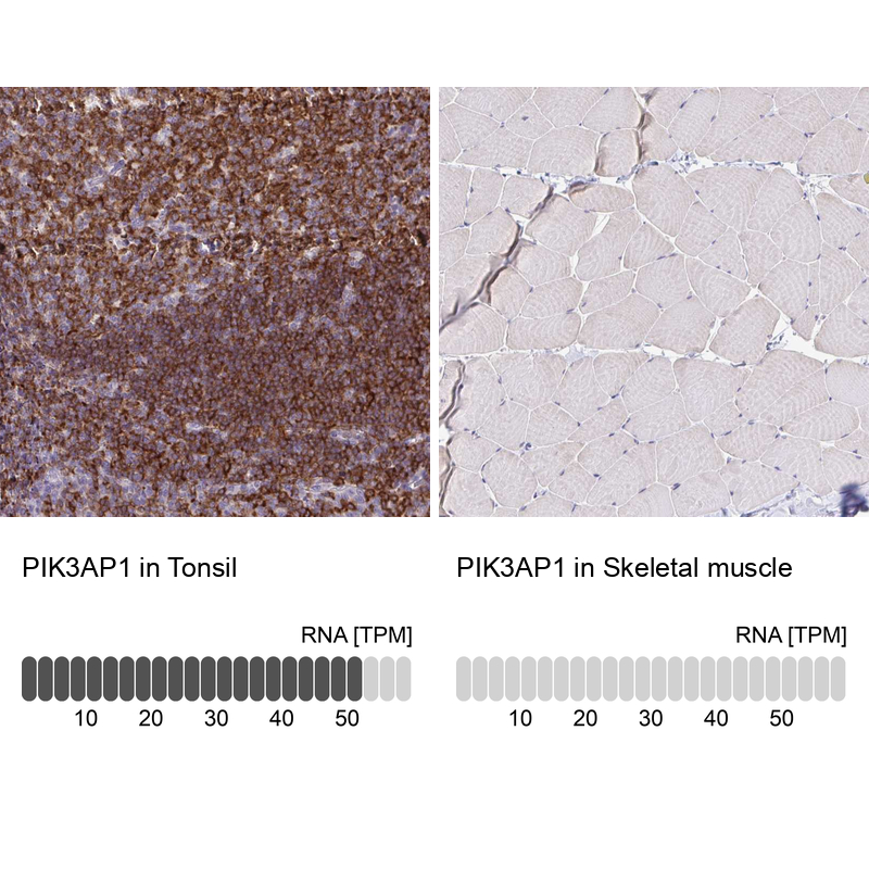
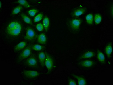
- Product NamePIK3AP1 Polyclonal Antibody
- Delivery Days Customer16
- ApplicationsImmunoFluorescence, Western Blot, ELISA, ImmunoCytoChemistry, ImmunoHistoChemistry, ImmunoHistoChemistry Frozen, ImmunoHistoChemistry Paraffin
- Applications SupplierWB(1:300-5000), ELISA(1:500-1000), IHC-P(1:200-400), IHC-F(1:100-500), IF(IHC-P)(1:50-200), IF(IHC-F)(1:50-200), IF(ICC)(1:50-200)
- CertificationResearch Use Only
- ClonalityPolyclonal
- Concentration1 ug/ul
- ConjugateUnconjugated
- HostRabbit
- IsotypeIgG
- ReactivityBovine, Canine, Equine, Human, Mouse, Rabbit, Rat, Sheep
- Storage Instruction-20°C
- UNSPSC41116161
Datasheet
Related products

Product group Antibodies
Anti-PIK3AP1 Antibody Picoband(r)A08114-CARRIER-FREE
ApplicationsWestern Blot, ELISA
ReactivityHuman
TargetPIK3AP1
- SizePrice
Product group Antibodies

BCAP / PIK3AP1 AntibodyLS-C674610
ApplicationsImmunoFluorescence, ELISA
ReactivityHuman
TargetPIK3AP1
- SizePrice
Product group Antibodies
Anti-PIK3AP1 AntibodyHPA038452
ApplicationsWestern Blot, ImmunoCytoChemistry, ImmunoHistoChemistry
ReactivityHuman
TargetPIK3AP1
- SizePrice
Product group Antibodies
PIK3AP1 AntibodyCSB-PA747806LA01HU
ApplicationsImmunoFluorescence, ELISA
ReactivityHuman
TargetPIK3AP1
- SizePrice
Product group Antibodies
PIK3AP1 Polyclonal AntibodyCAC13383
ApplicationsImmunoFluorescence, ELISA
TargetPIK3AP1
- SizePrice
Product group Antibodies
PIK3AP1 AntibodyPACO57232
ApplicationsImmunoFluorescence, ELISA
ReactivityHuman
TargetPIK3AP1
- SizePrice
![FACS analysis of HEK293T cells transfected with either PIK3AP1 plasmid(Red) or empty vector control plasmid(Blue) using GTX83893 PIK3AP1 antibody [7A11].](https://www.genetex.com/upload/website/prouct_img/normal/GTX83893/GTX83893_222_FACS_w_23061420_903.webp)
Product group Antibodies
PIK3AP1 antibody [7A11]GTX83893
ApplicationsFlow Cytometry, ImmunoFluorescence, Western Blot, ImmunoCytoChemistry, ImmunoHistoChemistry, ImmunoHistoChemistry Paraffin
ReactivityHuman
TargetPIK3AP1
- SizePrice