

PINK1 Antibody - N-terminal region (OAAB16695)
OAAB16695
ApplicationsWestern Blot, ImmunoHistoChemistry, ImmunoHistoChemistry Paraffin
Product group Antibodies
TargetPINK1
Overview
- SupplierAviva Systems Biology
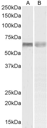
- Product NamePINK1 Antibody - N-terminal region (OAAB16695)
- Delivery Days Customer23
- ApplicationsWestern Blot, ImmunoHistoChemistry, ImmunoHistoChemistry Paraffin
- CertificationResearch Use Only
- ClonalityPolyclonal
- Concentration2 mg/ml
- FormatLiquid.PBS with 0.09% (W/V) sodium azide
- Gene ID65018
- Target namePINK1
- Target descriptionPTEN induced kinase 1
- Target synonymsBRPK; PARK6; protein kinase BRPK; PTEN induced putative kinase 1; PTEN-induced putative kinase protein 1; serine/threonine-protein kinase PINK1, mitochondrial
- HostRabbit
- Storage Instruction2°C to 8°C,-20°C
- UNSPSC12352203
Related products
Product group Antibodies

Goat anti-PINK1EB07940
ApplicationsFlow Cytometry, Western Blot, ELISA
TargetPINK1
- SizePrice
Product group Antibodies
Pink1 Polyclonal AntibodyCAC10977
ApplicationsELISA, ImmunoHistoChemistry
TargetPINK1
- SizePrice
Product group Antibodies
Anti-PINK1 Antibody144-66301
ApplicationsImmunoFluorescence, Western Blot
TargetPINK1
- SizePrice
Product group Antibodies
References
PINK1 antibody [N3C3]GTX107851
ApplicationsImmunoFluorescence, Western Blot, ImmunoCytoChemistry
TargetPINK1
- SizePrice

Product group Antibodies
Anti-PINK1 AntibodyHPA001931
ApplicationsImmunoHistoChemistry
TargetPINK1
- SizePrice
Product group Antibodies
Anti-PINK1 AntibodyA84378
ApplicationsFlow Cytometry, Western Blot, ELISA
- SizePrice
Product group Antibodies
PINK1 Polyclonal AntibodyBS-22173R
ApplicationsWestern Blot
TargetPINK1
- SizePrice

Product group Antibodies
Anti-PINK1 Antibody Picoband(r)A00201-2-CARRIER-FREE
ApplicationsFlow Cytometry, ImmunoFluorescence, Western Blot, ELISA, ImmunoCytoChemistry
TargetPINK1
- SizePrice