

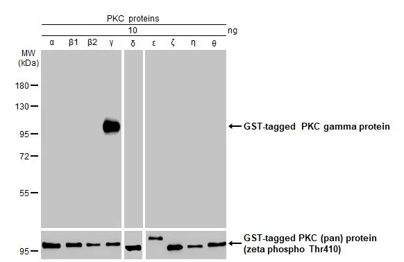
PKC proteins (10 ng) were separated by 7.5% SDS-PAGE, and the membrane was blotted with PKC gamma antibody [HL2226] (GTX638271) diluted at 1:1000. The HRP-conjugated anti-rabbit IgG antibody (GTX213110-01) was used to detect the primary antibody.
![PKC gamma antibody [HL2226] detects PKC gamma protein by immunohistochemical analysis. Sample: Paraffin-embedded rat tissues. PKC gamma stained by PKC gamma antibody [HL2226] (GTX638271) diluted at 1:100. Antigen Retrieval: Citrate buffer, pH 6.0, 15 min](https://www.genetex.com/upload/website/prouct_img/normal/GTX638271/GTX638271_T-44956_20230325_IHC-P_multiple_R_23032819_229.webp "PKC gamma antibody [HL2226] detects PKC gamma protein by immunohistochemical analysis. Sample: Paraffin-embedded rat tissues. PKC gamma stained by PKC gamma antibody [HL2226] (GTX638271) diluted at 1:100. Antigen Retrieval: Citrate buffer, pH 6.0, 15 min")
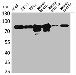
![Various tissue extracts (50 μg) were separated by 7.5% SDS-PAGE, and the membrane was blotted with PKC gamma antibody [HL2226] (GTX638271) diluted at 1:5000. The HRP-conjugated anti-rabbit IgG antibody (GTX213110-01) was used to detect the primary antibody.](https://www.genetex.com/upload/website/prouct_img/normal/GTX638271/GTX638271_45040_20230512_WB_M_R_23051702_478.webp "Various tissue extracts (50 μg) were separated by 7.5% SDS-PAGE, and the membrane was blotted with PKC gamma antibody [HL2226] (GTX638271) diluted at 1:5000. The HRP-conjugated anti-rabbit IgG antibody (GTX213110-01) was used to detect the primary antibody.")
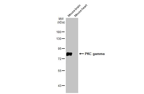
![Human brain (5 μg) was separated by 7.5% SDS-PAGE, and the membrane was blotted with PKC gamma antibody [HL2226] (GTX638271) diluted at 1:1000. The HRP-conjugated anti-rabbit IgG antibody (GTX213110-01) was used to detect the primary antibody.](https://www.genetex.com/upload/website/prouct_img/normal/GTX638271/GTX638271_45040_20230512_WB_brain_23051702_106.webp "Human brain (5 μg) was separated by 7.5% SDS-PAGE, and the membrane was blotted with PKC gamma antibody [HL2226] (GTX638271) diluted at 1:1000. The HRP-conjugated anti-rabbit IgG antibody (GTX213110-01) was used to detect the primary antibody.")
![PKC gamma antibody [HL2226] detects PKC gamma protein by immunohistochemical analysis. Sample: Paraffin-embedded mouse hippocampus. PKC gamma stained by PKC gamma antibody [HL2226] (GTX638271) diluted at 1:1000. Antigen Retrieval: Citrate buffer, pH 6.0, 15 min](https://www.genetex.com/upload/website/prouct_img/normal/GTX638271/GTX638271_45040_20251114_IHC-P_M_2_25112800_512.webp "PKC gamma antibody [HL2226] detects PKC gamma protein by immunohistochemical analysis. Sample: Paraffin-embedded mouse hippocampus. PKC gamma stained by PKC gamma antibody [HL2226] (GTX638271) diluted at 1:1000. Antigen Retrieval: Citrate buffer, pH 6.0, 15 min")
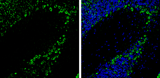
![PKC gamma antibody [HL2226] detects PKC gamma protein by immunohistochemical analysis. Sample: Paraffin-embedded rat cerebellum. Green: PKC gamma stained by PKC gamma antibody [HL2226] (GTX638271) diluted at 1:400. Red: beta Tubulin 3/ Tuj1 antibody [GT11710] (GTX631836) diluted at 1:500. Blue: Fluoroshield with DAPI (GTX30920). Antigen Retrieval: Citrate buffer, pH 6.0, 15 min](https://www.genetex.com/upload/website/prouct_img/normal/GTX638271/GTX638271_45040_20251121_IHC-P_R_2_25112800_499.webp "PKC gamma antibody [HL2226] detects PKC gamma protein by immunohistochemical analysis. Sample: Paraffin-embedded rat cerebellum. Green: PKC gamma stained by PKC gamma antibody [HL2226] (GTX638271) diluted at 1:400. Red: beta Tubulin 3/ Tuj1 antibody [GT11710] (GTX631836) diluted at 1:500. Blue: Fluoroshield with DAPI (GTX30920). Antigen Retrieval: Citrate buffer, pH 6.0, 15 min")
![PKC gamma antibody [HL2226] detects PKC gamma protein by immunohistochemical analysis. Sample: Paraffin-embedded mouse brain. PKC gamma stained by PKC gamma antibody [HL2226] (GTX638271) diluted at 1:1000. Antigen Retrieval: Citrate buffer, pH 6.0, 15 min](https://www.genetex.com/upload/website/prouct_img/normal/GTX638271/GTX638271_45040_20251114_IHC-P_M_1_25112800_792.webp "PKC gamma antibody [HL2226] detects PKC gamma protein by immunohistochemical analysis. Sample: Paraffin-embedded mouse brain. PKC gamma stained by PKC gamma antibody [HL2226] (GTX638271) diluted at 1:1000. Antigen Retrieval: Citrate buffer, pH 6.0, 15 min")
![PKC gamma antibody [HL2226] detects PKC gamma protein by immunohistochemical analysis. Sample: Paraffin-embedded rat hippocampus. Green: PKC gamma stained by PKC gamma antibody [HL2226] (GTX638271) diluted at 1:400. Red: beta Tubulin 3/ Tuj1 antibody [GT11710] (GTX631836) diluted at 1:500. Blue: Fluoroshield with DAPI (GTX30920). Antigen Retrieval: Citrate buffer, pH 6.0, 15 min](https://www.genetex.com/upload/website/prouct_img/normal/GTX638271/GTX638271_45040_20251121_IHC-P_R_25112800_527.webp "PKC gamma antibody [HL2226] detects PKC gamma protein by immunohistochemical analysis. Sample: Paraffin-embedded rat hippocampus. Green: PKC gamma stained by PKC gamma antibody [HL2226] (GTX638271) diluted at 1:400. Red: beta Tubulin 3/ Tuj1 antibody [GT11710] (GTX631836) diluted at 1:500. Blue: Fluoroshield with DAPI (GTX30920). Antigen Retrieval: Citrate buffer, pH 6.0, 15 min")
![PKC gamma antibody [HL2226] detects PKC gamma protein by immunohistochemical analysis. Sample: Paraffin-embedded mouse tissues. PKC gamma stained by PKC gamma antibody [HL2226] (GTX638271) diluted at 1:1000. Antigen Retrieval: Citrate buffer, pH 6.0, 15 min](https://www.genetex.com/upload/website/prouct_img/normal/GTX638271/GTX638271_45040_20251114_IHC-P_M_Multiple_RPKM_1_25112800_608.webp "PKC gamma antibody [HL2226] detects PKC gamma protein by immunohistochemical analysis. Sample: Paraffin-embedded mouse tissues. PKC gamma stained by PKC gamma antibody [HL2226] (GTX638271) diluted at 1:1000. Antigen Retrieval: Citrate buffer, pH 6.0, 15 min")
![PKC gamma antibody [HL2226] detects PKC gamma protein by immunohistochemical analysis. Sample: Paraffin-embedded mouse tissues. PKC gamma stained by PKC gamma antibody [HL2226] (GTX638271) diluted at 1:1000. Antigen Retrieval: Citrate buffer, pH 6.0, 15 min](https://www.genetex.com/upload/website/prouct_img/normal/GTX638271/GTX638271_45040_20251114_IHC-P_M_Multiple_RPKM_2_25112800_427.webp "PKC gamma antibody [HL2226] detects PKC gamma protein by immunohistochemical analysis. Sample: Paraffin-embedded mouse tissues. PKC gamma stained by PKC gamma antibody [HL2226] (GTX638271) diluted at 1:1000. Antigen Retrieval: Citrate buffer, pH 6.0, 15 min")
PKC proteins (10 ng) were separated by 7.5% SDS-PAGE, and the membrane was blotted with PKC gamma antibody [HL2226] (GTX638271) diluted at 1:1000. The HRP-conjugated anti-rabbit IgG antibody (GTX213110-01) was used to detect the primary antibody.
PKC gamma antibody [HL2226]
GTX638271
ApplicationsWestern Blot, ImmunoHistoChemistry, ImmunoHistoChemistry Paraffin
Product group Antibodies
ReactivityHuman, Mouse, Rat
TargetPRKCG
Overview
- SupplierGeneTex
- Product NamePKC gamma antibody [HL2226]
- Delivery Days Customer9
- Application Supplier NoteWB: 1:500-1:3000. *Optimal dilutions/concentrations should be determined by the researcher.Not tested in other applications.
- ApplicationsWestern Blot, ImmunoHistoChemistry, ImmunoHistoChemistry Paraffin
- CertificationResearch Use Only
- ClonalityMonoclonal
- Clone IDHL2226
- Concentration1 mg/ml
- ConjugateUnconjugated
- Gene ID5582
- Target namePRKCG
- Target descriptionprotein kinase C gamma
- Target synonymsPKC-gamma, PKCC, PKCG, PKCI(3), PKCgamma, SCA14, protein kinase C gamma type
- HostRabbit
- IsotypeIgG
- Protein IDP05129
- Protein NameProtein kinase C gamma type
- Scientific DescriptionProtein kinase C (PKC) is a family of serine- and threonine-specific protein kinases that can be activated by calcium and second messenger diacylglycerol. PKC family members phosphorylate a wide variety of protein targets and are known to be involved in diverse cellular signaling pathways. PKC also serve as major receptors for phorbol esters, a class of tumor promoters. Each member of the PKC family has a specific expression profile and is believed to play distinct roles in cells. The protein encoded by this gene is one of the PKC family members. This protein kinase is expressed solely in the brain and spinal cord and its localization is restricted to neurons. It has been demonstrated that several neuronal functions, including long term potentiation (LTP) and long term depression (LTD), specifically require this kinase. Knockout studies in mice also suggest that this kinase may be involved in neuropathic pain development. Defects in this protein have been associated with neurodegenerative disorder spinocerebellar ataxia-14 (SCA14). Two transcript variants encoding different isoforms have been found for this gene. [provided by RefSeq, Oct 2015]
- ReactivityHuman, Mouse, Rat
- Storage Instruction-20°C or -80°C,2°C to 8°C
- UNSPSC41116161
Datasheet
Related products
Product group Antibodies
PRKCG AntibodyCSB-PA004920
ApplicationsWestern Blot, ELISA
ReactivityHuman, Mouse, Rat
TargetPRKCG
- SizePrice

Product group Antibodies
Anti-PKC gamma/PRKCG Antibody Picoband(r)A01890-CARRIER-FREE
ApplicationsImmunoFluorescence, Western Blot, ImmunoCytoChemistry, ImmunoHistoChemistry
ReactivityHuman, Mouse, Rat
TargetPRKCG
- SizePrice
Product group Antibodies
Anti-PRKCG AntibodyA97329
ApplicationsWestern Blot, ELISA
ReactivityHuman, Mouse, Rat
- SizePrice

Product group Antibodies
Anti-PRKCG AntibodyHPA047870
ApplicationsImmunoHistoChemistry
ReactivityHuman
TargetPRKCG
- SizePrice
Product group Antibodies

PRKCG / PKC-Gamma AntibodyLS-C402929
ApplicationsWestern Blot, ELISA
ReactivityHuman, Mouse, Rat
TargetPRKCG
- SizePrice
![PKC proteins (10 ng) were separated by 7.5% SDS-PAGE, and the membrane was blotted with PKC gamma antibody [HL2227] (GTX638272) diluted at 1:1000. The HRP-conjugated anti-rabbit IgG antibody (GTX213110-01) was used to detect the primary antibody, and the signal was developed with Trident ECL plus-Enhanced.](https://www.genetex.com/upload/website/prouct_img/normal/GTX638272/GTX638272_T-44956_20230331_WB_Ag_23032819_813.webp)
Product group Antibodies
PKC gamma antibody [HL2227]GTX638272
ApplicationsImmunoFluorescence, Western Blot, ImmunoCytoChemistry
ReactivityHuman, Mouse, Rat
TargetPRKCG
- SizePrice
Product group Antibodies
PKC gamma antibodyGTX102539
ApplicationsWestern Blot, ImmunoHistoChemistry, ImmunoHistoChemistry Frozen
ReactivityHuman, Mouse, Rat
TargetPRKCG
- SizePrice
Product group Antibodies
PKC gamma antibodyGTX107639
ApplicationsWestern Blot, ImmunoHistoChemistry, ImmunoHistoChemistry Frozen, ImmunoHistoChemistry Paraffin
ReactivityHuman, Mouse, Rat
TargetPRKCG
- SizePrice