

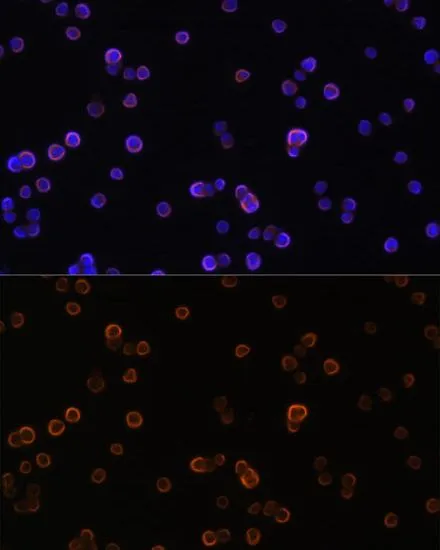
ICC/IF analysis of Raw264.7 cells using GTX32793 PLCG2 antibody. Blue : DAPI Dilution : 1:100


ICC/IF analysis of Raw264.7 cells using GTX32793 PLCG2 antibody. Blue : DAPI Dilution : 1:100
PLCG2 antibody
GTX32793
ApplicationsImmunoFluorescence, Western Blot, ImmunoCytoChemistry, ImmunoHistoChemistry, ImmunoHistoChemistry Paraffin
Product group Antibodies
ReactivityHuman, Mouse
TargetPLCG2
Overview
- SupplierGeneTex
- Product NamePLCG2 antibody
- Delivery Days Customer9
- Application Supplier NoteWB: 1:500 - 1:2000. ICC/IF: 1:20 - 1:100. IHC-P: 1:50 - 1:200. *Optimal dilutions/concentrations should be determined by the researcher.Not tested in other applications.
- ApplicationsImmunoFluorescence, Western Blot, ImmunoCytoChemistry, ImmunoHistoChemistry, ImmunoHistoChemistry Paraffin
- CertificationResearch Use Only
- ClonalityPolyclonal
- ConjugateUnconjugated
- Gene ID5336
- Target namePLCG2
- Target descriptionphospholipase C gamma 2
- Target synonymsAPLAID, FCAS3, PLC-IV, PLC-gamma-2, 1-phosphatidylinositol 4,5-bisphosphate phosphodiesterase gamma-2, phosphoinositide phospholipase C-gamma-2, phospholipase C, gamma 2 (phosphatidylinositol-specific), phospholipase C-IV
- HostRabbit
- IsotypeIgG
- Protein IDP16885
- Protein Name1-phosphatidylinositol 4,5-bisphosphate phosphodiesterase gamma-2
- Scientific DescriptionThe protein encoded by this gene is a transmembrane signaling enzyme that catalyzes the conversion of 1-phosphatidyl-1D-myo-inositol 4,5-bisphosphate to 1D-myo-inositol 1,4,5-trisphosphate (IP3) and diacylglycerol (DAG) using calcium as a cofactor. IP3 and DAG are second messenger molecules important for transmitting signals from growth factor receptors and immune system receptors across the cell membrane. Mutations in this gene have been found in autoinflammation, antibody deficiency, and immune dysregulation syndrome and familial cold autoinflammatory syndrome 3. [provided by RefSeq, Mar 2014]
- ReactivityHuman, Mouse
- Storage Instruction-20°C or -80°C,2°C to 8°C
- UNSPSC12352203
Datasheet
Related products
Product group Antibodies
Anti-PLCG2 Antibody144-02182
ApplicationsImmunoFluorescence, Western Blot, ImmunoHistoChemistry
ReactivityHuman, Mouse
TargetPLCG2
- SizePrice
Product group Antibodies
ApplicationsWestern Blot, ELISA, ImmunoCytoChemistry, ImmunoHistoChemistry, ImmunoHistoChemistry Frozen, ImmunoHistoChemistry Paraffin
ReactivityMouse, Rat
TargetPLCG2
- SizePrice
Product group Antibodies
PLCG 2 Recombinant AntibodyBSM-62254R
ApplicationsFlow Cytometry, ImmunoFluorescence, ImmunoPrecipitation, Western Blot, ImmunoHistoChemistry, ImmunoHistoChemistry Frozen, ImmunoHistoChemistry Paraffin
ReactivityHuman
TargetPLCG2
- SizePrice
Product group Antibodies
PLCG2 AntibodyCSB-PA010540
ApplicationsWestern Blot, ELISA, ImmunoHistoChemistry
ReactivityHuman, Mouse, Rat
TargetPLCG2
- SizePrice
Product group Antibodies
Anti-PLCG2 AntibodyA95807
ApplicationsWestern Blot, ELISA, ImmunoHistoChemistry
ReactivityHuman, Mouse, Rat
- SizePrice
Product group Antibodies
PLCG2 (phospho Tyr753) antibodyGTX32240
ApplicationsWestern Blot, ImmunoHistoChemistry, ImmunoHistoChemistry Paraffin
ReactivityHuman, Mouse, Rat
TargetPLCG2
- SizePrice
![Untreated (–) and treated (+) Raji whole cell extract (30 μg) were separated by 5% SDS-PAGE, and the membrane was blotted with PLCG2 (phospho Tyr759) antibody [HL3329] (GTX641094) diluted at 1:5000. The HRP-conjugated anti-rabbit IgG antibody (GTX213110-01) was used to detect the primary antibody, and the signal was developed with Trident ECL plus-Enhanced.](https://www.genetex.com/upload/website/prouct_img/normal/GTX641094/GTX641094_T-45551_20241011_WB_treatment_Pervanadate_24101600_226.webp)
Product group Antibodies
ApplicationsWestern Blot
ReactivityHuman
TargetPLCG2
- SizePrice
![Untreated (–) and treated (+) Raji whole cell extracts (30 μg) were separated by 5% SDS-PAGE, and the membrane was blotted with PLCG2 (phospho Tyr1217) antibody [HL3332] (GTX641097) diluted at 1:15000. The HRP-conjugated anti-rabbit IgG antibody (GTX213110-01) was used to detect the primary antibody.](https://www.genetex.com/upload/website/prouct_img/normal/GTX641097/GTX641097_T-45551_20241025_WB_treatment_PVD_24103022_414.webp)
Product group Antibodies
ApplicationsWestern Blot
ReactivityHuman
TargetPLCG2
- SizePrice
![Untreated (–) and treated (+) Raji whole cell extracts (30 μg) were separated by 5% SDS-PAGE, and the membrane was blotted with PLCG2 (phospho Tyr1217) antibody [HL3406] (GTX641243) diluted at 1:1000. The HRP-conjugated anti-rabbit IgG antibody (GTX213110-01) was used to detect the primary antibody.](https://www.genetex.com/upload/website/prouct_img/normal/GTX641243/GTX641243_T-45579_20241101_WB_treatment_pervanadate_24110700_328.webp)
Product group Antibodies
ApplicationsWestern Blot
ReactivityHuman
TargetPLCG2
- SizePrice