

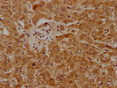
IHC image of CSB-PA018188LA01HU diluted at 1:1000 and staining in paraffin-embedded human liver tissue performed on a Leica BondTM system. After dewaxing and hydration, antigen retrieval was mediated by high pressure in a citrate buffer (pH 6.0). Section was blocked with 10% normal goat serum 30min at RT. Then primary antibody (1% BSA) was incubated at 4°C overnight. The primary is detected by a biotinylated secondary antibody and visualized using an HRP conjugated SP system.
IHC image of CSB-PA018188LA01HU diluted at 1:1000 and staining in paraffin-embedded human liver tissue performed on a Leica BondTM system. After dewaxing and hydration, antigen retrieval was mediated by high pressure in a citrate buffer (pH 6.0). Section was blocked with 10% normal goat serum 30min at RT. Then primary antibody (1% BSA) was incubated at 4°C overnight. The primary is detected by a biotinylated secondary antibody and visualized using an HRP conjugated SP system.
PLG Antibody
CSB-PA018188LA01HU
ApplicationsELISA, ImmunoHistoChemistry
Product group Antibodies
ReactivityHuman
TargetPLG
Overview
- SupplierCusabio
- Product NamePLG Antibody
- Delivery Days Customer20
- ApplicationsELISA, ImmunoHistoChemistry
- CertificationResearch Use Only
- ClonalityPolyclonal
- ConjugateUnconjugated
- Gene ID5340
- Target namePLG
- Target descriptionplasminogen
- Target synonymsHAE4, plasminogen, plasmin
- HostRabbit
- IsotypeIgG
- Protein IDP00747
- Protein NamePlasminogen
- Scientific DescriptionPlasmin dissolves the fibrin of blood clots and acts as a proteolytic factor in a variety of other processes including embryonic development, tissue remodeling, tumor invasion, and inflammation. In ovulation, weakens the walls of the Graafian follicle. It activates the urokinase-type plasminogen activator, collagenases and several complement zymogens, such as C1 and C5. Cleavage of fibronectin and laminin leads to cell detachment and apoptosis. Also cleaves fibrin, thrombospondin and von Willebrand factor. Its role in tissue remodeling and tumor invasion may be modulated by CSPG4. Binds to cells.
- ReactivityHuman
- Storage Instruction-20°C or -80°C
- UNSPSC41116161
Related products
Product group Antibodies
Anti-Plasminogen AntibodyA116764
ApplicationsDot Blot, ImmunoFluorescence, Western Blot, ELISA
ReactivityHuman
- SizePrice

Product group Antibodies
Anti-Plasminogen/PLG Antibody Picoband(r)A00702-1-CARRIER-FREE
ApplicationsWestern Blot, ELISA
ReactivityHuman, Mouse, Rat
TargetPLG
- SizePrice
Product group Antibodies
Anti-PLG Antibody144-03320
ApplicationsWestern Blot
ReactivityHuman, Rat
TargetPLG
- SizePrice
Product group Antibodies
Plasminogen Polyclonal AntibodyBS-1828R
ApplicationsImmunoFluorescence, Western Blot, ELISA, ImmunoHistoChemistry, ImmunoHistoChemistry Frozen, ImmunoHistoChemistry Paraffin
ReactivityBovine, Canine, Human, Mouse, Porcine, Rat
TargetPLG
- SizePrice
Product group Antibodies
ApplicationsImmunoPrecipitation, Western Blot, ImmunoCytoChemistry, ImmunoHistoChemistry
ReactivityPorcine, Rat
TargetPLG
- SizePrice

Product group Antibodies
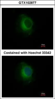
Plasminogen antibodyGTX102877
ApplicationsImmunoFluorescence, Western Blot, ImmunoCytoChemistry, ImmunoHistoChemistry, ImmunoHistoChemistry Paraffin
ReactivityHuman, Mouse, Rat
TargetPLG
- SizePrice
Product group Antibodies
Anti-PLG AntibodyHPA021602
ApplicationsWestern Blot, ImmunoHistoChemistry
ReactivityHuman
TargetPLG
- SizePrice
Product group Antibodies

ApplicationsWestern Blot, ELISA
ReactivityHuman
TargetPLG
- SizePrice