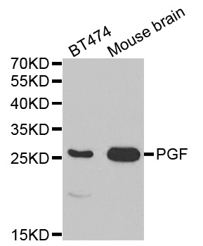
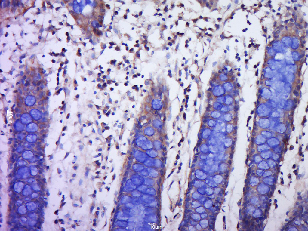
PLGF(PLGF94), CF405S conjugate, 0.1mg/mL [26628-22-8]
BNC040094
ReactivityBovine, Human, Mouse
Product group Antibodies
TargetPGF
Overview
- SupplierBiotium
- Product NamePLGF(PLGF94), CF405S conjugate, 0.1mg/mL [26628-22-8]
- Delivery Days Customer9
- CAS Number26628-22-8
- CertificationResearch Use Only
- ClonalityMonoclonal
- Clone IDPLGF94
- Concentration0.1 mg/ml
- ConjugateOther Conjugate
- Gene ID5228
- Target namePGF
- Target descriptionplacental growth factor
- Target synonymsD12S1900, PGFL, PIGF, PLGF, PlGF-2, SHGC-10760, placenta growth factor, placental growth factor, vascular endothelial growth factor-related protein
- HostMouse
- IsotypeIgG1
- Protein IDP49763
- Protein NamePlacenta growth factor
- Scientific DescriptionThe onset of angiogenesis is believed to be an early event in tumorigenesis and may facilitate tumor progression and metastasis. Several growth factors with angiogenic activity have been described. These include Fibroblast Growth Factor (FGF), Platelet Derived Growth Factor (PDGF), Vascular Endothelial Growth Factor (VEGF) and Placenta Growth Factor (PLGF). Placenta growth factor (PLGF) is a secreted protein primarily produced by placental trophoblasts but also expressed in other endothelial cells and tumors. There are three isoforms, PLGF-1, PLGF-2, and PLGF-3. PLGF-2 is expressed up until week 8 in the placenta; the placental tissues continuously express PLGF-1 and PLGF-3 but only PLGF-1 is found in colon and mammary carcinomas. PLGF acts to stimulate angiogenesis, endothelial growth and migration. PLGF is a powerful promoter of tumor growth and is upregulated in some cancers, and PLGF is thought to aid in atherosclerotic lesions and neovascular growth surrounding the lesion. Also, PLGF appears to aid aldosterone mediated atherosclerosis. Serum levels of PLGF in some cases are used as a potential biomarker for disease or genetic defect. Recent research indicates that PLGF levels are lower in mothers with Down syndrome fetuses. Evidence has suggested VEGF to be an obligatory component in PLGF signaling. While VEGF homodimers and VEGF/PLGF heterodimers function as potent mediators of mitogenic and chemotactic responses in endothelial cells, PLGF homodimers are effectual only at extremely high concentrations. Indeed, many of the physiological effects attributed to VEGF may actually be a result of VEGF/PLGF. VEGF and PLGF share a common receptor, Flt-1, and may also activate Flk-1/KDR.Primary antibodies are available purified, or with a selection of fluorescent CF® Dyes and other labels. CF® Dyes offer exceptional brightness and photostability. Note: Conjugates of blue fluorescent dyes like CF®405S and CF®405M are not recommended for detecting low abundance targets, because blue dyes have lower fluorescence and can give higher non-specific background than other dye colors.
- SourceAnimal
- ReactivityBovine, Human, Mouse
- Storage Instruction2°C to 8°C,RT
- UNSPSC41116161


![Sandwich ELISA detection of recombinant Human PLGF protein, His tag (GTX138450-pro) using antibodies as below. Capture: PLGF antibody [GT57] (GTX641446) (5 μg/mL) Detection: PLGF antibody [HL2682] (GTX639346) (1 μg/mL)](https://www.genetex.com/upload/website/prouct_img/normal/GTX639346/GTX639346_T-45222_20241220_ELISA_PAIR_24123021_606.webp)

