

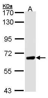
Sample (30 ug of whole cell lysate) A: H1299 7.5% SDS PAGE GTX102537 diluted at 1:1000
Sample (30 ug of whole cell lysate) A: H1299 7.5% SDS PAGE GTX102537 diluted at 1:1000
PPEF1 antibody [N1N2], N-term
GTX102537
ApplicationsWestern Blot
Product group Antibodies
ReactivityHuman
TargetPPEF1
Overview
- SupplierGeneTex
- Product NamePPEF1 antibody [N1N2], N-term
- Delivery Days Customer9
- Application Supplier NoteWB: 1:500-1:3000. *Optimal dilutions/concentrations should be determined by the researcher.Not tested in other applications.
- ApplicationsWestern Blot
- CertificationResearch Use Only
- ClonalityPolyclonal
- Concentration0.12 mg/ml
- ConjugateUnconjugated
- Gene ID5475
- Target namePPEF1
- Target descriptionprotein phosphatase with EF-hand domain 1
- Target synonymsPP7, PPEF, PPP7C, PPP7CA, serine/threonine-protein phosphatase with EF-hands 1, protein phosphatase 7, catalytic subunit, alpha isozyme, protein phosphatase with EF calcium-binding domain, protein phosphatase, EF-hand calcium binding domain 1, protein phosphatase, serine/threonine type, with EF-hands, serine/threonine protein phosphatase 7
- HostRabbit
- IsotypeIgG
- Protein IDO14829
- Protein NameSerine/threonine-protein phosphatase with EF-hands 1
- Scientific DescriptionThis gene encodes a member of the serine/threonine protein phosphatase with EF-hand motif family. The protein contains a protein phosphatase catalytic domain, and at least two EF-hand calcium-binding motifs in its C terminus. Although its substrate(s) is unknown, the encoded protein has been suggested to play a role in specific sensory neuron function and/or development. This gene shares high sequence similarity with the Drosophila retinal degeneration C (rdgC) gene. Several alternatively spliced transcript variants, each encoding a distinct isoform, have been described. [provided by RefSeq]
- ReactivityHuman
- Storage Instruction-20°C or -80°C,2°C to 8°C
- UNSPSC41116161
Datasheet
Related products
Product group Antibodies
PPEF1 AntibodyCSB-PA018435LA01HU
ApplicationsWestern Blot, ELISA, ImmunoHistoChemistry
ReactivityHuman, Mouse
TargetPPEF1
- SizePrice

Product group Antibodies
Anti-PPEF1 Antibody Picoband(r)A11074-1-CARRIER-FREE
ApplicationsFlow Cytometry, ImmunoFluorescence, Western Blot, ELISA, ImmunoCytoChemistry
ReactivityHuman
TargetPPEF1
- SizePrice
Product group Antibodies

PPEF / PPEF1 AntibodyLS-C830842
ApplicationsWestern Blot, ELISA, ImmunoHistoChemistry
ReactivityHuman
TargetPPEF1
- SizePrice

Product group Antibodies
Anti-PPEF1 AntibodyHPA034577
ApplicationsImmunoHistoChemistry
ReactivityHuman
TargetPPEF1
- SizePrice
Product group Antibodies
Ppef1 Polyclonal AntibodyCAC08745
ApplicationsWestern Blot, ELISA, ImmunoHistoChemistry
ReactivityMouse
TargetPPEF1
- SizePrice
Product group Antibodies
PPEF1 antibodyGTX114652
ApplicationsWestern Blot, ImmunoHistoChemistry, ImmunoHistoChemistry Paraffin
ReactivityHuman
TargetPPEF1
- SizePrice
Product group Antibodies
Anti-PPEF1 (Center) Antibody102-21849
ApplicationsWestern Blot
TargetPPEF1
- SizePrice