Bio-Connect

Primary

373 to 384 of 587 products
Sort by
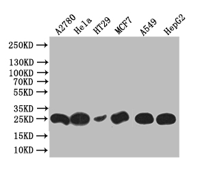
Western Blot Positive WB detected in: HT29 whole cell lysate, Hela whole cell lysate, HT29 whole cell lysate, MCF7 whole cell lysate, A549 whole cell lysate, HEPG2 whole cell lysate All lanes: HE4 antibody at 1:1000 Secondary Goat polyclonal to rabbit IgG at 1/50000 dilution Predicted band size: 25 kDa Observed band size: 25 kDa
Product group Antibodies
Cusabio
ApplicationsFlow Cytometry, Western Blot, ELISA
ReactivityHuman
TargetWFDC2
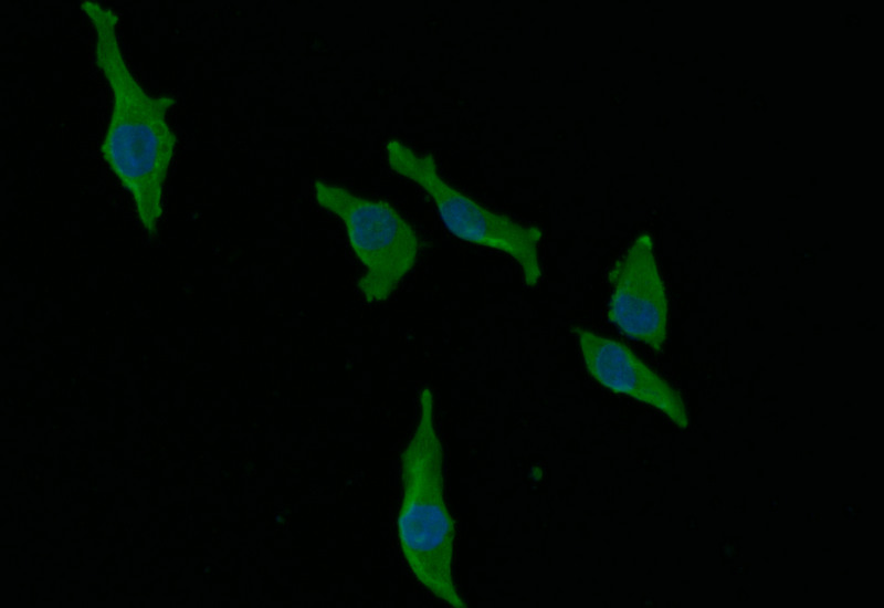
Immunofluorescence staining of MCF-7 with CSB-RA965823A0HU at 1:30, counter-stained with DAPI. The cells were fixed in 4% formaldehyde and blocked in 10% normal Goat Serum. The cells were then incubated with the antibody overnight at 4°C. The secondary antibody was Alexa Fluor 492-congugated AffiniPure Goat Anti-Rabbit IgG(H+L).
Product group Antibodies
Cusabio
ApplicationsFlow Cytometry, ImmunoFluorescence, ELISA
ReactivityHuman
TargetAGO2
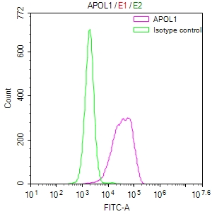
Overlay Peak curve showing HepG2 cells stained with CSB-RA966671A0HU (red line) at 1:50. The cells were fixed in 4% formaldehyde and permeated by 0.2% TritonX-100. Then 10% normal goat serum to block non-specific protein-protein interactions followed by the antibody (1microg/1*106cells) for 45min at 4°C. The secondary antibody used was FITC-conjugated Goat Anti-rabbit IgG(H+L) at 1:200 dilution for 35min at 4°C.Control antibody (green line) was rabbit IgG (1microg/1*106cells) used under the same conditions. Acquisition of >10,000 events was performed.
Product group Antibodies
Cusabio
ApplicationsFlow Cytometry, ELISA
ReactivityHuman
TargetAPOL1
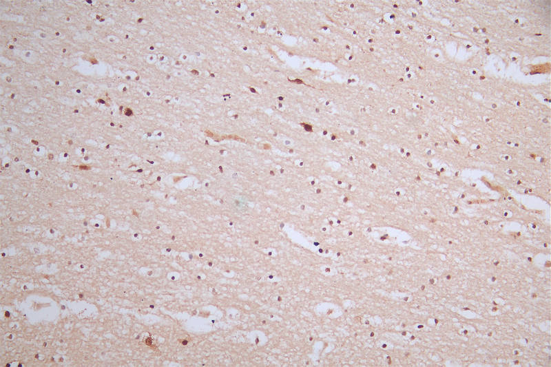
IHC image of CSB-RA980583A0HU diluted at 1:50 and staining in paraffin-embedded human brain tissue performed on a Leica BondTM system. After dewaxing and hydration, antigen retrieval was mediated by high pressure in a citrate buffer (pH 6.0). Section was blocked with 10% normal goat serum 30min at RT. Then primary antibody (1% BSA) was incubated at 4°C overnight. The primary is detected by a Goat anti-rabbit polymer IgG labeled by HRP and visualized using 0.57% DAB.
Product group Antibodies
Cusabio
ApplicationsFlow Cytometry, ImmunoFluorescence, ELISA, ImmunoHistoChemistry
ReactivityHuman
TargetMDM2
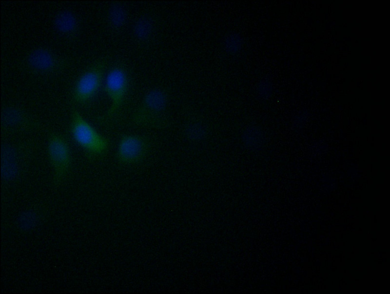
Immunofluorescence staining of A549 with CSB-RA982003A0HU at 1:60, counter-stained with DAPI. The cells were fixed in 4% formaldehyde and blocked in 10% normal Goat Serum. The cells were then incubated with the antibody overnight at 4°C. The secondary antibody was Alexa Fluor 522-congugated AffiniPure Goat Anti-Rabbit IgG(H+L).
Product group Antibodies
Cusabio
ApplicationsFlow Cytometry, ImmunoFluorescence, ELISA
ReactivityHuman
TargetNEUROD1
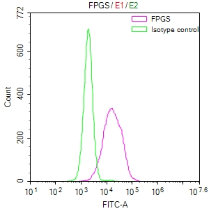
Overlay Peak curve showing HepG2 cells stained with CSB-RA983477A0HU (red line) at 1:50. The cells were fixed in 4% formaldehyde and permeated by 0.2% TritonX-100. Then 10% normal goat serum to block non-specific protein-protein interactions followed by the antibody (1microg/1*106cells) for 45min at 4°C. The secondary antibody used was FITC-conjugated Goat Anti-rabbit IgG(H+L) at 1:200 dilution for 35min at 4°C.Control antibody (green line) was rabbit IgG (1microg/1*106cells) used under the same conditions. Acquisition of >10,000 events was performed.
Product group Antibodies
Cusabio
ApplicationsFlow Cytometry, ELISA
ReactivityHuman
TargetFPGS
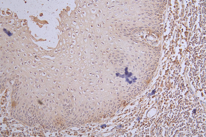
IHC image of CSB-RA984483A0HU diluted at 1:100 and staining in paraffin-embedded human tonsil tissue performed on a Leica BondTM system. After dewaxing and hydration, antigen retrieval was mediated by high pressure in a citrate buffer (pH 6.0). Section was blocked with 10% normal goat serum 30min at RT. Then primary antibody (1% BSA) was incubated at 4°C overnight. The primary is detected by a Goat anti-rabbit polymer IgG labeled by HRP and visualized using 0.65% DAB.
Product group Antibodies
Cusabio
ApplicationsFlow Cytometry, ELISA, ImmunoHistoChemistry
ReactivityHuman
TargetMKI67
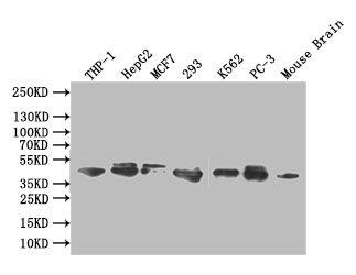
Western Blot Positive WB detected in: THP-1 whole cell lysate, HEPG2 whole cell lysate,MCF7 whole cell lysate,293 whole cell lysate,K562 whole cell lysate,PC-3 whole cell lysate, Mouse brain tissue lysate All lanes: ERK1/2 antibody at 1:1000 Secondary Goat polyclonal to rabbit IgG at 1/50000 dilution Predicted band size: 42,44 kDa Observed band size: 42,44 kDa
Product group Antibodies
Cusabio
ApplicationsFlow Cytometry, Western Blot, ELISA, ImmunoHistoChemistry
ReactivityHuman, Mouse
TargetMAPK1
Immunofluorescence staining of HepG2 with CSB-RA986776A0HU at 1:50, counter-stained with DAPI. The cells were fixed in 4% formaldehyde and blocked in 10% normal Goat Serum. The cells were then incubated with the antibody overnight at 4°C. The secondary antibody was Alexa Fluor 510-congugated AffiniPure Goat Anti-Rabbit IgG(H+L).
Product group Antibodies
Cusabio
ApplicationsFlow Cytometry, ImmunoFluorescence, ELISA
ReactivityHuman
TargetGSN
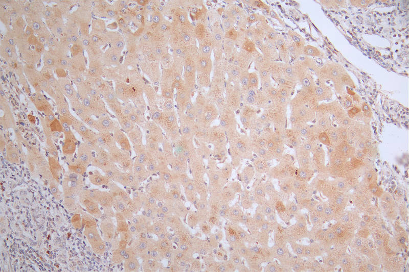
IHC image of CSB-RA988767A0HU diluted at 1:50 and staining in paraffin-embedded human liver cancer performed on a Leica BondTM system. After dewaxing and hydration, antigen retrieval was mediated by high pressure in a citrate buffer (pH 6.0). Section was blocked with 10% normal goat serum 30min at RT. Then primary antibody (1% BSA) was incubated at 4°C overnight. The primary is detected by a Goat anti-rabbit polymer IgG labeled by HRP and visualized using 0.38% DAB.
Product group Antibodies
Cusabio
ApplicationsELISA, ImmunoHistoChemistry
ReactivityHuman
TargetCRP
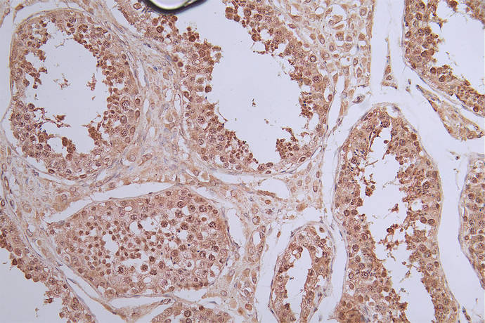
IHC image of CSB-RA989713A0HU diluted at 1:50 and staining in paraffin-embedded human testis tissue performed on a Leica BondTM system. After dewaxing and hydration, antigen retrieval was mediated by high pressure in a citrate buffer (pH 6.0). Section was blocked with 10% normal goat serum 30min at RT. Then primary antibody (1% BSA) was incubated at 4°C overnight. The primary is detected by a Goat anti-rabbit polymer IgG labeled by HRP and visualized using 0.40% DAB.
Product group Antibodies
Cusabio
ApplicationsFlow Cytometry, ImmunoFluorescence, ELISA, ImmunoHistoChemistry
ReactivityHuman
TargetSTAT1
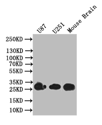
Western Blot Positive WB detected in: U87 whole cell lysate, U251 whole cell lysate, Mouse Brain tissue lysate All lanes: BDNF antibody at 1:1000 Secondary Goat polyclonal to rabbit IgG at 1/50000 dilution Predicted band size: 28 kDa Observed band size: 28 kDa
Product group Antibodies
Cusabio
ApplicationsWestern Blot, ELISA
ReactivityHuman, Mouse
TargetBDNF