

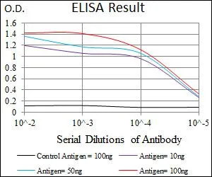
ELISA analysis of antigen using GTX60673 PSAP antibody [4D5F4]. Black : Control antigen 100ng Purple : Antigen 10ng Blue : Antigen 50ng Red : Antigen 100ng
![FACS analysis of HeLa cells using GTX60673 PSAP antibody [4D5F4]. Green : PSAP Purple : negative control](https://www.genetex.com/upload/website/prouct_img/normal/GTX60673/GTX60673_20170912_FACS_w_23061123_308.webp "FACS analysis of HeLa cells using GTX60673 PSAP antibody [4D5F4]. Green : PSAP Purple : negative control")
![ICC/IF analysis of HepG2 cells using GTX60673 PSAP antibody [4D5F4]. Green : PSAP Blue: DRAQ5 fluorescent DNA dye Red: Actin filaments](https://www.genetex.com/upload/website/prouct_img/normal/GTX60673/GTX60673_20170912_ICCIF_w_23061123_914.webp "ICC/IF analysis of HepG2 cells using GTX60673 PSAP antibody [4D5F4]. Green : PSAP Blue: DRAQ5 fluorescent DNA dye Red: Actin filaments")
![IHC-P analysis of human pancreas tissue using GTX60673 PSAP antibody [4D5F4].](https://www.genetex.com/upload/website/prouct_img/normal/GTX60673/GTX60673_20170912_IHC-P_w_23061123_824.webp "IHC-P analysis of human pancreas tissue using GTX60673 PSAP antibody [4D5F4].")
![WB analysis of HEK293 (1) and PSAP (AA: 325-524)-hIgGFc transfected HEK293 (2) cell lysate using GTX60673 PSAP antibody [4D5F4].](https://www.genetex.com/upload/website/prouct_img/normal/GTX60673/GTX60673_20170912_WB_w_23061123_831.webp "WB analysis of HEK293 (1) and PSAP (AA: 325-524)-hIgGFc transfected HEK293 (2) cell lysate using GTX60673 PSAP antibody [4D5F4].")
ELISA analysis of antigen using GTX60673 PSAP antibody [4D5F4]. Black : Control antigen 100ng Purple : Antigen 10ng Blue : Antigen 50ng Red : Antigen 100ng
PSAP antibody [4D5F4]
GTX60673
ApplicationsFlow Cytometry, ImmunoFluorescence, Western Blot, ELISA, ImmunoCytoChemistry, ImmunoHistoChemistry, ImmunoHistoChemistry Paraffin
Product group Antibodies
ReactivityHuman
TargetPSAP
Overview
- SupplierGeneTex
- Product NamePSAP antibody [4D5F4]
- Delivery Days Customer9
- Application Supplier NoteWB: 1/500 - 1/2000. ICC/IF: 1/50-1/500. IHC-P: 1/200 - 1/1000. FACS: 1/200 - 1/400. ELISA: 1/10000. *Optimal dilutions/concentrations should be determined by the researcher.Not tested in other applications.
- ApplicationsFlow Cytometry, ImmunoFluorescence, Western Blot, ELISA, ImmunoCytoChemistry, ImmunoHistoChemistry, ImmunoHistoChemistry Paraffin
- CertificationResearch Use Only
- ClonalityMonoclonal
- Clone ID4D5F4
- Concentration1 mg/ml
- ConjugateUnconjugated
- Gene ID5660
- Target namePSAP
- Target descriptionprosaposin
- Target synonymsGLBA, PARK24, PSAPD, SAP1, SAP2, prosaposin, precursor of saposins, proactivator polypeptide, saposin-A, saposin-B, saposin-C, saposin-D, sphingolipid activator protein-1, sphingolipid activator protein-2
- HostMouse
- IsotypeIgG1
- Protein IDP07602
- Protein NameProsaposin
- Scientific DescriptionThis gene encodes a highly conserved glycoprotein which is a precursor for 4 cleavage products: saposins A, B, C, and D. Each domain of the precursor protein is approximately 80 amino acid residues long with nearly identical placement of cysteine residues and glycosylation sites. Saposins A-D localize primarily to the lysosomal compartment where they facilitate the catabolism of glycosphingolipids with short oligosaccharide groups. The precursor protein exists both as a secretory protein and as an integral membrane protein and has neurotrophic activities. Mutations in this gene have been associated with Gaucher disease, Tay-Sachs disease, and metachromatic leukodystrophy. Alternative splicing results in multiple transcript variants encoding different isoforms. [provided by RefSeq, Jul 2008]
- ReactivityHuman
- Storage Instruction-20°C or -80°C,2°C to 8°C
- UNSPSC12352203
Datasheet
Related products
Product group Antibodies
PSAP Polyclonal AntibodyCAC13883
ApplicationsImmunoFluorescence, Western Blot, ELISA, ImmunoHistoChemistry
ReactivityMouse
TargetPSAP
- SizePrice
Product group Antibodies
Anti-PSAP Antibody118-10033
ApplicationsELISA, ImmunoHistoChemistry
ReactivityHuman
- SizePrice
Product group Antibodies
Anti-PSAP AntibodyA101324
ApplicationsWestern Blot, ELISA
ReactivityHuman
- SizePrice
Product group Antibodies
References
PSAP antibody [N1N3]GTX101064
ApplicationsWestern Blot, ImmunoHistoChemistry, ImmunoHistoChemistry Frozen, ImmunoHistoChemistry Paraffin
ReactivityHuman, Mouse, Rat
TargetPSAP
- SizePrice
Product group Antibodies
PSAP antibody [N3C3]GTX101065
ApplicationsImmunoHistoChemistry, ImmunoHistoChemistry Paraffin
ReactivityHuman
TargetPSAP
- SizePrice
Product group Antibodies
PSAP Polyclonal AntibodyBS-1879R
ApplicationsImmunoFluorescence, Western Blot, ELISA, ImmunoCytoChemistry, ImmunoHistoChemistry, ImmunoHistoChemistry Frozen, ImmunoHistoChemistry Paraffin
ReactivityCanine, Human, Mouse, Rat
TargetPSAP
- SizePrice
Product group Antibodies
PSAP antibodyGTX54581
ApplicationsImmunoFluorescence, Western Blot, ImmunoCytoChemistry, ImmunoHistoChemistry, ImmunoHistoChemistry Paraffin
ReactivityHuman, Mouse, Rat
TargetPSAP
- SizePrice
Product group Antibodies

PSAP / Prosaposin AntibodyLS-C331707
ApplicationsImmunoFluorescence, Western Blot, ImmunoHistoChemistry
ReactivityHuman, Mouse, Rat
TargetPSAP
- SizePrice