

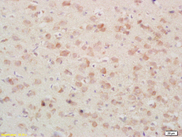
Formalin-fixed and paraffin embedded rat brain tissue labeled Anti-PSAP/PAP Polyclonal Antibody, Unconjugated (bs-1879R) at 1:200, followed by conjugation to the secondary antibody and DAB staining
 at 1:1000 dilution and 4˚C overnight incubation. Followed by conjugated secondary antibody incubation at 1:20000 for 60 min at 37˚C")
Formalin-fixed and paraffin embedded rat brain tissue labeled Anti-PSAP/PAP Polyclonal Antibody, Unconjugated (bs-1879R) at 1:200, followed by conjugation to the secondary antibody and DAB staining
PSAP Polyclonal Antibody
BS-1879R
ApplicationsImmunoFluorescence, Western Blot, ELISA, ImmunoCytoChemistry, ImmunoHistoChemistry, ImmunoHistoChemistry Frozen, ImmunoHistoChemistry Paraffin
Product group Antibodies
ReactivityCanine, Human, Mouse, Rat
TargetPSAP
Overview
- SupplierBioss
- Product NamePSAP Polyclonal Antibody
- Delivery Days Customer16
- ApplicationsImmunoFluorescence, Western Blot, ELISA, ImmunoCytoChemistry, ImmunoHistoChemistry, ImmunoHistoChemistry Frozen, ImmunoHistoChemistry Paraffin
- Applications SupplierWB(1:300-5000), ELISA(1:500-1000), IHC-P(1:200-400), IHC-F(1:100-500), IF(IHC-P)(1:50-200), IF(IHC-F)(1:50-200), IF(ICC)(1:50-200)
- CertificationResearch Use Only
- ClonalityPolyclonal
- Concentration1 ug/ul
- ConjugateUnconjugated
- Gene ID5660
- Target namePSAP
- Target descriptionprosaposin
- Target synonymsGLBA, PARK24, PSAPD, SAP1, SAP2, prosaposin, precursor of saposins, proactivator polypeptide, saposin-A, saposin-B, saposin-C, saposin-D, sphingolipid activator protein-1, sphingolipid activator protein-2
- HostRabbit
- IsotypeIgG
- Protein IDP07602
- Protein NameProsaposin
- ReactivityCanine, Human, Mouse, Rat
- Storage Instruction-20°C
- UNSPSC41116161
Datasheet
Related products
Product group Antibodies
Anti-PSAP Antibody118-10033
ApplicationsELISA, ImmunoHistoChemistry
ReactivityHuman
- SizePrice
Product group Antibodies
Anti-PSAP AntibodyA101324
ApplicationsWestern Blot, ELISA
ReactivityHuman
- SizePrice

Product group Antibodies
Anti-PSAP Antibody Picoband(r)A00937-1-CARRIER-FREE
ApplicationsFlow Cytometry, ImmunoFluorescence, Western Blot, ELISA, ImmunoCytoChemistry, ImmunoHistoChemistry
ReactivityHuman
TargetPSAP
- SizePrice
Product group Antibodies
PSAP Polyclonal AntibodyCAC13883
ApplicationsImmunoFluorescence, Western Blot, ELISA, ImmunoHistoChemistry
ReactivityMouse
TargetPSAP
- SizePrice
Product group Antibodies
PSAP AntibodyCSB-PA018836DA01HU
ApplicationsImmunoFluorescence, Western Blot, ELISA, ImmunoHistoChemistry
ReactivityHuman, Mouse
TargetPSAP
- SizePrice
![Wild-type (WT) and PSAP knockout (KO) HeLa cell extracts (30 μg) were separated by 10% SDS-PAGE, and the membrane was blotted with PSAP antibody [N1N3] (GTX101064) diluted at 1:500. The HRP-conjugated anti-rabbit IgG antibody (GTX213110-01) was used to detect the primary antibody.](https://www.genetex.com/upload/website/prouct_img/normal/GTX101064/GTX101064_40142_20181109_WB_KO_watermark_w_23060100_816.webp)
Product group Antibodies
PSAP antibody [N1N3]GTX101064
ApplicationsWestern Blot, ImmunoHistoChemistry, ImmunoHistoChemistry Frozen, ImmunoHistoChemistry Paraffin
ReactivityHuman, Mouse, Rat
TargetPSAP
- SizePrice
Product group Antibodies

PSAP / Prosaposin AntibodyLS-C331707
ApplicationsImmunoFluorescence, Western Blot, ImmunoHistoChemistry
ReactivityHuman, Mouse, Rat
TargetPSAP
- SizePrice

Product group Antibodies
Anti-PSAP AntibodyHPA004426
ApplicationsWestern Blot, ImmunoCytoChemistry, ImmunoHistoChemistry
ReactivityHuman
TargetPSAP
- SizePrice
Product group Antibodies
Anti-MRPL40 AntibodyCAB18191
ApplicationsWestern Blot, ELISA
ReactivityHuman
TargetPSAP
- SizePrice