

PSAP / Prosaposin Antibody (clone 4LJ, Ready-to-Use)
LS-C170843
ApplicationsImmunoHistoChemistry, ImmunoHistoChemistry Frozen, ImmunoHistoChemistry Paraffin
Product group Antibodies
TargetPSAP
Overview
- SupplierLifeSpan BioSciences
- Product NamePSAP / Prosaposin Antibody (clone 4LJ, Ready-to-Use)
- Delivery Days Customer23
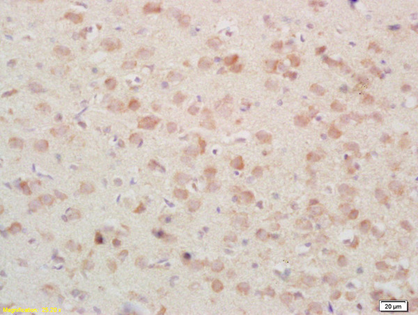
- Application Supplier NoteIHC: The prediluted antibody can be used on frozen cryostat sections as well as formalin-fixed paraffinembedded tissue sections. For paraffin-embedded tissue sections, we recommend an incubation time and temperature of 30 minutes at 37°C for this antibody, when used in conjunction with immunoperoxidase staining kit. Prolonged fixation in buffered formalin can destroy the epitope. The applications listed have been tested for the unmodified form of this product. Other forms have not been tested.. IHC, IHC-Fr, IHC-P IHC: The prediluted antibody can be used on frozen cryostat sections as well as formalin-fixed paraffinembedded tissue sections. For paraffin-embedded tissue sections, we recommend an incubation time and temperature of 30 minutes at 37°C for this antibody, when used in conjunction with immunoperoxidase staining kit. Prolonged fixation in buffered formalin can destroy the epitope. The applications listed have been tested for the unmodified form of this product. Other forms have not been tested.
- ApplicationsImmunoHistoChemistry, ImmunoHistoChemistry Frozen, ImmunoHistoChemistry Paraffin
- CertificationResearch Use Only
- ClonalityMonoclonal
- Clone ID4LJ
- ConjugateUnconjugated
- Estimated Purity...
- Gene ID5660
- Target namePSAP
- Target descriptionprosaposin
- Target synonymsGLBA; PARK24; proactivator polypeptide; prosaposin; PSAPD; SAP1; SAP2; saposin-A; saposin-B; saposin-C; saposin-D; sphingolipid activator protein-1; sphingolipid activator protein-2
- HostMouse
- IsotypeIgG2a
- Storage Instruction-20°C,2°C to 8°C
- UNSPSC12352203
Related products
Product group Antibodies
PSAP Polyclonal AntibodyBS-1879R
ApplicationsImmunoFluorescence, Western Blot, ELISA, ImmunoCytoChemistry, ImmunoHistoChemistry, ImmunoHistoChemistry Frozen, ImmunoHistoChemistry Paraffin
TargetPSAP
- SizePrice
Product group Antibodies
PSAP Polyclonal AntibodyCAC13883
ApplicationsImmunoFluorescence, Western Blot, ELISA, ImmunoHistoChemistry
TargetPSAP
- SizePrice
Product group Antibodies
PSAP AntibodyCSB-PA018836DA01HU
ApplicationsImmunoFluorescence, Western Blot, ELISA, ImmunoHistoChemistry
ReactivityHuman, Mouse
TargetPSAP
- SizePrice

Product group Antibodies
Anti-PSAP AntibodyHPA004426
ApplicationsWestern Blot, ImmunoCytoChemistry, ImmunoHistoChemistry
TargetPSAP
- SizePrice
Product group Antibodies

PSAP / Prosaposin AntibodyLS-C331707
ApplicationsImmunoFluorescence, Western Blot, ImmunoHistoChemistry
TargetPSAP
- SizePrice

Product group Antibodies
Anti-PSAP Antibody Picoband(r)A00937-1-CARRIER-FREE
ApplicationsFlow Cytometry, ImmunoFluorescence, Western Blot, ELISA, ImmunoCytoChemistry, ImmunoHistoChemistry
TargetPSAP
- SizePrice