

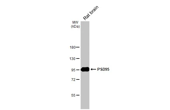
Rat tissue extract (50 μg) was separated by 7.5% SDS-PAGE, and the membrane was blotted with PSD95 antibody [HL2382] (GTX638590) diluted at 1:1000. The HRP-conjugated anti-rabbit IgG antibody (GTX213110-01) was used to detect the primary antibody.
![Non-transfected (–) and transfected (+) 293T whole cell extracts (5 μg) were separated by 7.5% SDS-PAGE, and the membrane was blotted with PSD95 antibody [HL2382] (GTX638590) diluted at 1:5000. The HRP-conjugated anti-rabbit IgG antibody (GTX213110-01) was used to detect the primary antibody.](https://www.genetex.com/upload/website/prouct_img/normal/GTX638590/GTX638590_T-45040_20240621_WB_B_24062501_536.webp "Non-transfected (–) and transfected (+) 293T whole cell extracts (5 μg) were separated by 7.5% SDS-PAGE, and the membrane was blotted with PSD95 antibody [HL2382] (GTX638590) diluted at 1:5000. The HRP-conjugated anti-rabbit IgG antibody (GTX213110-01) was used to detect the primary antibody.")
![PSD95 antibody [HL2382] detects PSD95 protein at synapse by immunofluorescent analysis. Sample: DIV9 rat E18 primary hippocampal neuron cells were fixed in 4% paraformaldehyde at RT for 15 min. Green: PSD95 stained by PSD95 antibody [HL2382] (GTX638590) diluted at 1:250. Red: Tau, an axon marker, stained by Tau antibody [GT287] (GTX634809) diluted at 1:500. Blue: Fluoroshield with DAPI (GTX30920).](https://www.genetex.com/upload/website/prouct_img/normal/GTX638590/GTX638590_T-45040_20230519_ICC_IF_R_24072519_396.webp "PSD95 antibody [HL2382] detects PSD95 protein at synapse by immunofluorescent analysis. Sample: DIV9 rat E18 primary hippocampal neuron cells were fixed in 4% paraformaldehyde at RT for 15 min. Green: PSD95 stained by PSD95 antibody [HL2382] (GTX638590) diluted at 1:250. Red: Tau, an axon marker, stained by Tau antibody [GT287] (GTX634809) diluted at 1:500. Blue: Fluoroshield with DAPI (GTX30920).")
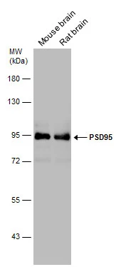
![Various tissue extracts (50 μg) were separated by 7.5% SDS-PAGE, and the membrane was blotted with PSD95 antibody [HL2382] (GTX638590) diluted at 1:1000. The HRP-conjugated anti-rabbit IgG antibody (GTX213110-01) was used to detect the primary antibody.](https://www.genetex.com/upload/website/prouct_img/normal/GTX638590/GTX638590_45502_20240830_WB_M_tissue_24090423_396.webp "Various tissue extracts (50 μg) were separated by 7.5% SDS-PAGE, and the membrane was blotted with PSD95 antibody [HL2382] (GTX638590) diluted at 1:1000. The HRP-conjugated anti-rabbit IgG antibody (GTX213110-01) was used to detect the primary antibody.")
Rat tissue extract (50 μg) was separated by 7.5% SDS-PAGE, and the membrane was blotted with PSD95 antibody [HL2382] (GTX638590) diluted at 1:1000. The HRP-conjugated anti-rabbit IgG antibody (GTX213110-01) was used to detect the primary antibody.
PSD95 antibody [HL2382]
GTX638590
ApplicationsImmunoFluorescence, Western Blot, ImmunoCytoChemistry
Product group Antibodies
ReactivityHuman, Mouse, Rat
TargetDlg4
Overview
- SupplierGeneTex
- Product NamePSD95 antibody [HL2382]
- Delivery Days Customer7
- Application Supplier NoteWB: 1:500-1:10000. *Optimal dilutions/concentrations should be determined by the researcher.Not tested in other applications.
- ApplicationsImmunoFluorescence, Western Blot, ImmunoCytoChemistry
- CertificationResearch Use Only
- ClonalityMonoclonal
- Clone IDHL2382
- Concentration1 mg/ml
- ConjugateUnconjugated
- Gene ID13385
- Target nameDlg4
- Target descriptiondiscs large MAGUK scaffold protein 4
- Target synonymsDlgh4, PSD-95, PSD95, SAP90, SAP90A, disks large homolog 4, PSD-95 alpha 2b, PSD-95 beta, SAP-90, discs large homolog 4, discs, large homolog 4, postsynaptic density protein 95, synapse-associated protein 90
- HostRabbit
- IsotypeIgG
- Protein IDQ62108
- Protein NameDisks large homolog 4
- Scientific DescriptionEnables ionotropic glutamate receptor binding activity and scaffold protein binding activity. Involved in several processes, including dendritic spine morphogenesis; locomotory exploration behavior; and neurotransmitter receptor localization to postsynaptic specialization membrane. Acts upstream of or within protein localization to synapse; regulation of long-term neuronal synaptic plasticity; and synaptic vesicle maturation. Located in several cellular components, including axon; postsynaptic density membrane; and synaptic vesicle. Is extrinsic component of cytoplasmic side of plasma membrane. Part of AMPA glutamate receptor complex. Is active in glutamatergic synapse. Colocalizes with postsynaptic membrane. Is expressed in central nervous system; dorsal root ganglion; olfactory epithelium; and retina. Used to study Williams-Beuren syndrome and autism spectrum disorder. Human ortholog(s) of this gene implicated in autosomal dominant non-syndromic intellectual disability. Orthologous to human DLG4 (discs large MAGUK scaffold protein 4). [provided by Alliance of Genome Resources, Apr 2022]
- ReactivityHuman, Mouse, Rat
- Storage Instruction-20°C or -80°C,2°C to 8°C
- UNSPSC41116161
Datasheet
Related products
Product group Antibodies
PSD95 Recombinant AntibodyBSM-60886R
ApplicationsImmunoFluorescence, Western Blot, ImmunoHistoChemistry, ImmunoHistoChemistry Frozen, ImmunoHistoChemistry Paraffin
ReactivityMouse, Rat
TargetDlg4
- SizePrice
Product group Antibodies
ApplicationsImmunoPrecipitation, Western Blot, ImmunoCytoChemistry, ImmunoHistoChemistry
ReactivityMouse
TargetDlg4
- SizePrice
![PSD95 antibody detects PSD95 protein at nucleus by immunohistochemical analysis. Sample: Paraffin-embedded mouse eye. Green: PSD95 stained by PSD95 antibody (GTX133091) diluted at 1:500. Red: beta Tubulin 3/ Tuj1 , a Cytoskeleton marker, stained by beta Tubulin 3/ Tuj1 antibody [GT11710] (GTX631836) diluted at 1:500. Blue: Fluoroshield with DAPI (GTX30920). Antigen Retrieval: Citrate buffer, pH 6.0, 15 min](https://www.genetex.com/upload/website/prouct_img/normal/GTX133091/GTX133091_44615_20220624_IHC-P_M_22062919_492.webp)
Product group Antibodies
PSD95 antibodyGTX133091
ApplicationsImmunoFluorescence, Western Blot, ImmunoCytoChemistry, ImmunoHistoChemistry, ImmunoHistoChemistry Frozen, ImmunoHistoChemistry Paraffin
ReactivityHuman, Mouse, Rat
TargetDlg4
- SizePrice
Product group Antibodies
PSD95 antibodyGTX133167
ApplicationsImmunoFluorescence, Western Blot, ImmunoCytoChemistry, ImmunoHistoChemistry, ImmunoHistoChemistry Frozen, ImmunoHistoChemistry Paraffin
ReactivityMouse, Rat
TargetDlg4
- SizePrice
![PSD95 antibody detects PSD95 protein by immunohistochemical analysis. Samples: Paraffin-embedded mouse retina. Green: PSD95 protein stained by PSD95 antibody (GTX133255) diluted at 1:250. Red: beta Tubulin 3/ Tuj1, a marker, stained by beta Tubulin 3/ Tuj1 antibody [GT1338] (GTX631831) diluted at 1:500. Blue: Fluoroshield with DAPI (GTX30920).
Antigen Retrieval: Citrate buffer, pH 6.0, 15 min](https://www.genetex.com/upload/website/prouct_img/normal/GTX133255/GTX133255_42613_20171127_IHC-P_M_w_23060523_909.webp)
Product group Antibodies
PSD95 antibodyGTX133255
ApplicationsImmunoFluorescence, Western Blot, ImmunoCytoChemistry, ImmunoHistoChemistry, ImmunoHistoChemistry Frozen, ImmunoHistoChemistry Paraffin
ReactivityMouse, Rat
TargetDlg4
- SizePrice
![Mouse tissue extract (50 μg) was separated by 7.5% SDS-PAGE, and the membrane was blotted with PSD95 antibody [GT1436] (GTX634290) diluted at 1:1000. The HRP-conjugated anti-mouse IgG antibody (GTX213111-01) was used to detect the primary antibody.](https://www.genetex.com/upload/website/prouct_img/normal/GTX634290/GTX634290_42912_20180323_WB_M_brain_w_23061202_396.webp)
Product group Antibodies
PSD95 antibody [GT1436]GTX634290
ApplicationsWestern Blot
ReactivityMouse, Rat
TargetDlg4
- SizePrice
![Mouse tissue extract (50 μg) was separated by 7.5% SDS-PAGE, and the membrane was blotted with PSD95 antibody [GT1234] (GTX634291) diluted at 1:5000. The HRP-conjugated anti-mouse IgG antibody (GTX213111-01) was used to detect the primary antibody.](https://www.genetex.com/upload/website/prouct_img/normal/GTX634291/GTX634291_42912_20180323_WB_M_brain_w_23061202_830.webp)
Product group Antibodies
PSD95 antibody [GT1234]GTX634291
ApplicationsImmunoFluorescence, Western Blot, ImmunoCytoChemistry
ReactivityMouse, Rat
TargetDlg4
- SizePrice