

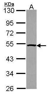
Sample (30 ug of whole cell lysate) A: JurKat 10% SDS PAGE GTX114675 diluted at 1:1000
 antibody at 1:200 dilution.")
 dilution: 1:500.
Antigen Retrieval: Trilogy? (EDTA based, pH 8.0) buffer, 15min")
Sample (30 ug of whole cell lysate) A: JurKat 10% SDS PAGE GTX114675 diluted at 1:1000
PSMC4 antibody
GTX114675
ApplicationsImmunoFluorescence, Western Blot, ImmunoCytoChemistry, ImmunoHistoChemistry, ImmunoHistoChemistry Paraffin
Product group Antibodies
ReactivityHuman
TargetPSMC4
Overview
- SupplierGeneTex
- Product NamePSMC4 antibody
- Delivery Days Customer9
- Application Supplier NoteWB: 1:500-1:3000. ICC/IF: 1:100-1:1000. IHC-P: 1:100-1:1000. *Optimal dilutions/concentrations should be determined by the researcher.Not tested in other applications.
- ApplicationsImmunoFluorescence, Western Blot, ImmunoCytoChemistry, ImmunoHistoChemistry, ImmunoHistoChemistry Paraffin
- CertificationResearch Use Only
- ClonalityPolyclonal
- Concentration0.71 mg/ml
- ConjugateUnconjugated
- Gene ID5704
- Target namePSMC4
- Target descriptionproteasome 26S subunit, ATPase 4
- Target synonymsMIP224, RPT3, S6, TBP-7, TBP7, 26S proteasome regulatory subunit 6B, 26S protease regulatory subunit 6B, 26S proteasome AAA-ATPase subunit RPT3, MB67-interacting protein, Tat-binding protein 7, protease 26S subunit 6, proteasome (prosome, macropain) 26S subunit, ATPase, 4
- HostRabbit
- IsotypeIgG
- Protein IDP43686
- Protein Name26S proteasome regulatory subunit 6B
- Scientific DescriptionThe 26S proteasome is a multicatalytic proteinase complex with a highly ordered structure composed of 2 complexes, a 20S core and a 19S regulator. The 20S core is composed of 4 rings of 28 non-identical subunits; 2 rings are composed of 7 alpha subunits and 2 rings are composed of 7 beta subunits. The 19S regulator is composed of a base, which contains 6 ATPase subunits and 2 non-ATPase subunits, and a lid, which contains up to 10 non-ATPase subunits. Proteasomes are distributed throughout eukaryotic cells at a high concentration and cleave peptides in an ATP/ubiquitin-dependent process in a non-lysosomal pathway. An essential function of a modified proteasome, the immunoproteasome, is the processing of class I MHC peptides. This gene encodes one of the ATPase subunits, a member of the triple-A family of ATPases which have a chaperone-like activity. This subunit has been shown to interact with an orphan member of the nuclear hormone receptor superfamily highly expressed in liver, and with gankyrin, a liver oncoprotein. Two transcript variants encoding different isoforms have been identified. [provided by RefSeq]
- ReactivityHuman
- Storage Instruction-20°C or -80°C,2°C to 8°C
- UNSPSC41116161
Datasheet
Related products
Product group Antibodies
PSMC4 AntibodyCSB-PA018894LA01HU
ApplicationsWestern Blot, ELISA, ImmunoHistoChemistry
ReactivityHuman, Mouse
TargetPSMC4
- SizePrice
Product group Antibodies
Anti-PSMC4 Antibody144-02505
ApplicationsImmunoFluorescence, Western Blot
ReactivityHuman, Mouse
TargetPSMC4
- SizePrice

Product group Antibodies
Anti-PSMC4 Antibody Picoband(r)A06099-1-CARRIER-FREE
ApplicationsFlow Cytometry, ImmunoFluorescence, ImmunoPrecipitation, Western Blot, ELISA, ImmunoCytoChemistry, ImmunoHistoChemistry
ReactivityHuman, Mouse, Rat
TargetPSMC4
- SizePrice
Product group Antibodies
Anti-PSMC4 AntibodyA30566
ApplicationsImmunoFluorescence, Western Blot, ImmunoHistoChemistry
ReactivityHuman, Mouse, Rat
- SizePrice

Product group Antibodies
Anti-PSMC4 AntibodyHPA002044
ApplicationsWestern Blot, ImmunoCytoChemistry, ImmunoHistoChemistry
ReactivityHuman, Mouse, Rat
TargetPSMC4
- SizePrice
Product group Antibodies

PSMC4 AntibodyLS-C332127
ApplicationsImmunoFluorescence, Western Blot, ImmunoHistoChemistry
ReactivityHuman, Mouse
TargetPSMC4
- SizePrice
Product group Antibodies
PSMC4 Polyclonal AntibodyCAC14873
ApplicationsWestern Blot, ELISA, ImmunoHistoChemistry
ReactivityMouse
TargetPSMC4
- SizePrice
Product group Antibodies
PSMC4 antibodyGTX54612
ApplicationsImmunoFluorescence, Western Blot, ImmunoCytoChemistry
ReactivityHuman, Mouse
TargetPSMC4
- SizePrice
Product group Antibodies
TBP7 Recombinant AntibodyBSM-62823R
ApplicationsImmunoFluorescence, Western Blot, ImmunoCytoChemistry
ReactivityHuman, Mouse, Rat
TargetPSMC4
- SizePrice