

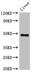
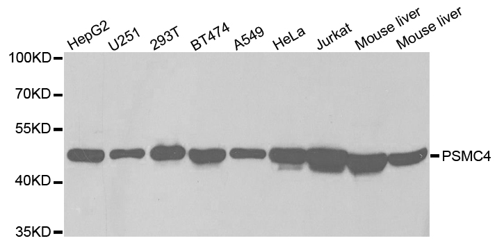
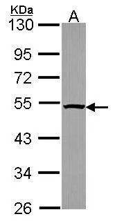
Western Blot Positive WB detected in: Mouse liver tissue All lanes: PSMC4 antibody at 3.4ug/ml Secondary Goat polyclonal to rabbit IgG at 1/50000 dilution Predicted band size: 48, 44 kDa Observed band size: 48 kDa
. Section was blocked with 10% normal goat serum 30min at RT. Then primary antibody (1% BSA) was incubated at 4°C overnight. The primary is detected by a biotinylated secondary antibody and visualized using an HRP conjugated SP system.")
. Section was blocked with 10% normal goat serum 30min at RT. Then primary antibody (1% BSA) was incubated at 4°C overnight. The primary is detected by a biotinylated secondary antibody and visualized using an HRP conjugated SP system.")
Western Blot Positive WB detected in: Mouse liver tissue All lanes: PSMC4 antibody at 3.4ug/ml Secondary Goat polyclonal to rabbit IgG at 1/50000 dilution Predicted band size: 48, 44 kDa Observed band size: 48 kDa
PSMC4 Antibody
CSB-PA018894LA01HU
ApplicationsWestern Blot, ELISA, ImmunoHistoChemistry
Product group Antibodies
ReactivityHuman, Mouse
TargetPSMC4
Overview
- SupplierCusabio
- Product NamePSMC4 Antibody
- Delivery Days Customer20
- ApplicationsWestern Blot, ELISA, ImmunoHistoChemistry
- CertificationResearch Use Only
- ClonalityPolyclonal
- ConjugateUnconjugated
- Gene ID5704
- Target namePSMC4
- Target descriptionproteasome 26S subunit, ATPase 4
- Target synonymsMIP224, RPT3, S6, TBP-7, TBP7, 26S proteasome regulatory subunit 6B, 26S protease regulatory subunit 6B, 26S proteasome AAA-ATPase subunit RPT3, MB67-interacting protein, Tat-binding protein 7, protease 26S subunit 6, proteasome (prosome, macropain) 26S subunit, ATPase, 4
- HostRabbit
- IsotypeIgG
- Protein IDP43686
- Protein Name26S proteasome regulatory subunit 6B
- Scientific DescriptionComponent of the 26S proteasome, a multiprotein complex involved in the ATP-dependent degradation of ubiquitinated proteins. This complex plays a key role in the maintenance of protein homeostasis by removing misfolded or damaged proteins, which could impair cellular functions, and by removing proteins whose functions are no longer required. Therefore, the proteasome participates in numerous cellular processes, including cell cycle progression, apoptosis, or DNA damage repair. PSMC4 belongs to the heterohexameric ring of AAA (ATPases associated with diverse cellular activities) proteins that unfolds ubiquitinated target proteins that are concurrently translocated into a proteolytic chamber and degraded into peptides.
- ReactivityHuman, Mouse
- Storage Instruction-20°C or -80°C
- UNSPSC41116161
Related products
Product group Antibodies
Anti-PSMC4 Antibody144-02505
ApplicationsImmunoFluorescence, Western Blot
ReactivityHuman, Mouse
TargetPSMC4
- SizePrice

Product group Antibodies
Anti-PSMC4 Antibody Picoband(r)A06099-1-CARRIER-FREE
ApplicationsFlow Cytometry, ImmunoFluorescence, ImmunoPrecipitation, Western Blot, ELISA, ImmunoCytoChemistry, ImmunoHistoChemistry
ReactivityHuman, Mouse, Rat
TargetPSMC4
- SizePrice
Product group Antibodies
Anti-PSMC4 AntibodyA30566
ApplicationsImmunoFluorescence, Western Blot, ImmunoHistoChemistry
ReactivityHuman, Mouse, Rat
- SizePrice

Product group Antibodies
Anti-PSMC4 AntibodyHPA002044
ApplicationsWestern Blot, ImmunoCytoChemistry, ImmunoHistoChemistry
ReactivityHuman, Mouse, Rat
TargetPSMC4
- SizePrice
Product group Antibodies

PSMC4 AntibodyLS-C332127
ApplicationsImmunoFluorescence, Western Blot, ImmunoHistoChemistry
ReactivityHuman, Mouse
TargetPSMC4
- SizePrice
Product group Antibodies
PSMC4 Polyclonal AntibodyCAC14873
ApplicationsWestern Blot, ELISA, ImmunoHistoChemistry
ReactivityMouse
TargetPSMC4
- SizePrice
Product group Antibodies
PSMC4 antibodyGTX114675
ApplicationsImmunoFluorescence, Western Blot, ImmunoCytoChemistry, ImmunoHistoChemistry, ImmunoHistoChemistry Paraffin
ReactivityHuman
TargetPSMC4
- SizePrice
Product group Antibodies
TBP7 Recombinant AntibodyBSM-62823R
ApplicationsImmunoFluorescence, Western Blot, ImmunoCytoChemistry
ReactivityHuman, Mouse, Rat
TargetPSMC4
- SizePrice