

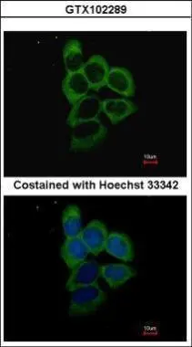
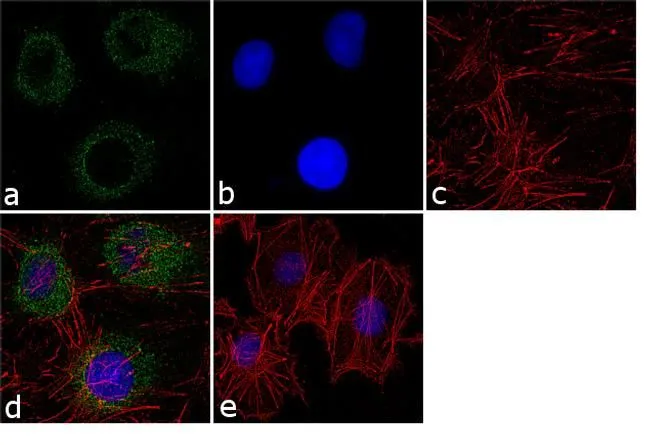
Immunofluorescence analysis of methanol-fixed A431, using PSMD2(GTX102289) antibody at 1:500 dilution.
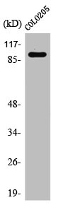
 A: JC B: BCL-1 7.5% SDS PAGE GTX102289 diluted at 1:1000 The HRP-conjugated anti-rabbit IgG antibody (GTX213110-01) was used to detect the primary antibody.")
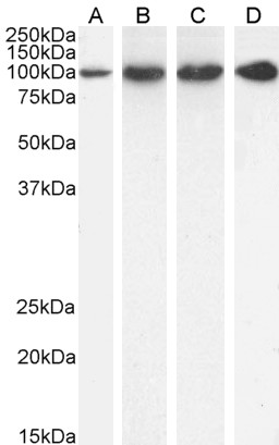
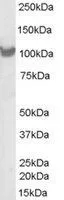
 A: A431 5% SDS PAGE GTX102289 diluted at 1:500 The HRP-conjugated anti-rabbit IgG antibody (GTX213110-01) was used to detect the primary antibody.")
Immunofluorescence analysis of methanol-fixed A431, using PSMD2(GTX102289) antibody at 1:500 dilution.
PSMD2 antibody
GTX102289
ApplicationsImmunoFluorescence, Western Blot, ImmunoCytoChemistry
Product group Antibodies
ReactivityHuman, Mouse
TargetPSMD2
Overview
- SupplierGeneTex
- Product NamePSMD2 antibody
- Delivery Days Customer9
- Application Supplier NoteWB: 1:500-1:3000. ICC/IF: 1:100-1:1000. *Optimal dilutions/concentrations should be determined by the researcher.Not tested in other applications.
- ApplicationsImmunoFluorescence, Western Blot, ImmunoCytoChemistry
- CertificationResearch Use Only
- ClonalityPolyclonal
- Concentration0.82 mg/ml
- ConjugateUnconjugated
- Gene ID5708
- Target namePSMD2
- Target descriptionproteasome 26S subunit ubiquitin receptor, non-ATPase 2
- Target synonymsP97, RPN1, S2, TRAP2, 26S proteasome non-ATPase regulatory subunit 2, 55.11 protein, TNFR-associated protein 2, proteasome (prosome, macropain) 26S subunit, non-ATPase, 2, proteasome 26S subunit, non-ATPase 2, protein 55.11, tumor necrosis factor type 1 receptor-associated protein 2
- HostRabbit
- IsotypeIgG
- Protein IDQ13200
- Protein Name26S proteasome non-ATPase regulatory subunit 2
- Scientific DescriptionThe 26S proteasome is a multicatalytic proteinase complex with a highly ordered structure composed of 2 complexes, a 20S core and a 19S regulator. The 20S core is composed of 4 rings of 28 non-identical subunits; 2 rings are composed of 7 alpha subunits and 2 rings are composed of 7 beta subunits. The 19S regulator is composed of a base, which contains 6 ATPase subunits and 2 non-ATPase subunits, and a lid, which contains up to 10 non-ATPase subunits. Proteasomes are distributed throughout eukaryotic cells at a high concentration and cleave peptides in an ATP/ubiquitin-dependent process in a non-lysosomal pathway. An essential function of a modified proteasome, the immunoproteasome, is the processing of class I MHC peptides. This gene encodes one of the non-ATPase subunits of the 19S regulator lid. In addition to participation in proteasome function, this subunit may also participate in the TNF signalling pathway since it interacts with the tumor necrosis factor type 1 receptor. A pseudogene has been identified on chromosome 1. [provided by RefSeq]
- ReactivityHuman, Mouse
- Storage Instruction-20°C or -80°C,2°C to 8°C
- UNSPSC41116161
Datasheet
Related products
Product group Antibodies
ApplicationsFlow Cytometry, ImmunoFluorescence, Western Blot, ELISA
ReactivityHuman, Mouse, Rat
- SizePrice

Product group Antibodies
Anti-PSMD2 Antibody Picoband(r)A06642-3-CARRIER-FREE
ApplicationsImmunoFluorescence, Western Blot, ELISA, ImmunoCytoChemistry
ReactivityHuman, Monkey, Rat
TargetPSMD2
- SizePrice
Product group Antibodies
Anti-PSMD2 Antibody144-01999
ApplicationsImmunoFluorescence, Western Blot, ImmunoHistoChemistry
ReactivityHuman, Mouse
TargetPSMD2
- SizePrice
Product group Antibodies
ApplicationsImmunoFluorescence, Western Blot, ImmunoHistoChemistry, ImmunoHistoChemistry Paraffin
ReactivityBovine, Canine, Chicken, Equine, Human, Mouse, Porcine, Rabbit, Rat, Sheep
TargetPSMD2
- SizePrice
Product group Antibodies
PSMD2 AntibodyCSB-PA003869
ApplicationsWestern Blot, ELISA, ImmunoHistoChemistry
ReactivityHuman, Mouse, Rat
TargetPSMD2
- SizePrice
Product group Antibodies

ApplicationsFlow Cytometry, ImmunoFluorescence, Western Blot, ELISA
ReactivityBovine, Canine, Human, Mouse, Rat
TargetPSMD2
- SizePrice
Product group Antibodies
Psmd2 Polyclonal AntibodyCAC09285
ApplicationsImmunoFluorescence, Western Blot, ELISA, ImmunoHistoChemistry
TargetPSMD2
- SizePrice
Product group Antibodies

PSMD2 AntibodyLS-C331815
ApplicationsImmunoFluorescence, Western Blot, ImmunoHistoChemistry
ReactivityHuman, Mouse
TargetPSMD2
- SizePrice
Product group Antibodies
PSMD2 antibodyGTX79499
ApplicationsImmunoFluorescence, Western Blot, ImmunoCytoChemistry
ReactivityCanine, Hamster, Human, Monkey, Mouse, Primate, Rat
TargetPSMD2
- SizePrice
Product group Antibodies
PSMD2 antibody, C-termGTX89633
ApplicationsWestern Blot
ReactivityHuman
TargetPSMD2
- SizePrice