

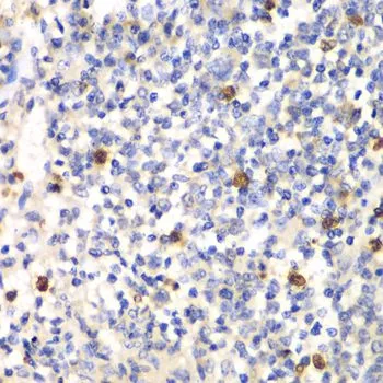
IHC-P analysis of mouse spleen tissue using GTX54625 PSMD7 antibody. Dilution : 1:100


IHC-P analysis of mouse spleen tissue using GTX54625 PSMD7 antibody. Dilution : 1:100
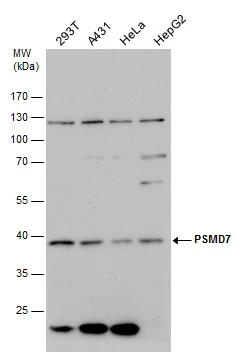
PSMD7 antibody
GTX54625
ApplicationsImmunoFluorescence, Western Blot, ImmunoCytoChemistry, ImmunoHistoChemistry, ImmunoHistoChemistry Paraffin
Product group Antibodies
ReactivityHuman, Mouse
TargetPSMD7
Overview
- SupplierGeneTex
- Product NamePSMD7 antibody
- Delivery Days Customer7
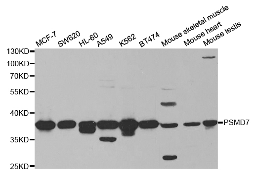
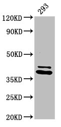
- Application Supplier NoteWB: 1:500 - 1:2000. ICC/IF: 1:50 - 1:200. IHC-P: 1:50 - 1:200. *Optimal dilutions/concentrations should be determined by the researcher.Not tested in other applications.
- ApplicationsImmunoFluorescence, Western Blot, ImmunoCytoChemistry, ImmunoHistoChemistry, ImmunoHistoChemistry Paraffin
- CertificationResearch Use Only
- ClonalityPolyclonal
- ConjugateUnconjugated
- Gene ID5713
- Target namePSMD7
- Target descriptionproteasome 26S subunit, non-ATPase 7
- Target synonymsMOV34, P40, Rpn8, S12, 26S proteasome non-ATPase regulatory subunit 7, 26S proteasome regulatory subunit S12, 26S proteasome regulatory subunit rpn8, Moloney leukemia virus-34 proviral integration, Mov34 homolog, proteasome (prosome, macropain) 26S subunit, non-ATPase, 7 (Mov34 homolog), proteasome subunit p40
- HostRabbit
- IsotypeIgG
- Protein IDP51665
- Protein Name26S proteasome non-ATPase regulatory subunit 7
- Scientific DescriptionThe 26S proteasome is a multicatalytic proteinase complex with a highly ordered structure composed of 2 complexes, a 20S core and a 19S regulator. The 20S core is composed of 4 rings of 28 non-identical subunits; 2 rings are composed of 7 alpha subunits and 2 rings are composed of 7 beta subunits. The 19S regulator is composed of a base, which contains 6 ATPase subunits and 2 non-ATPase subunits, and a lid, which contains up to 10 non-ATPase subunits. Proteasomes are distributed throughout eukaryotic cells at a high concentration and cleave peptides in an ATP/ubiquitin-dependent process in a non-lysosomal pathway. An essential function of a modified proteasome, the immunoproteasome, is the processing of class I MHC peptides. This gene encodes a non-ATPase subunit of the 19S regulator. A pseudogene has been identified on chromosome 17. [provided by RefSeq, Jul 2008]
- ReactivityHuman, Mouse
- Storage Instruction-20°C or -80°C,2°C to 8°C
- UNSPSC41116161
Datasheet
Related products
Product group Antibodies
Anti-PSMD7 AntibodyA30752
ApplicationsImmunoFluorescence, Western Blot, ImmunoHistoChemistry
ReactivityHuman, Mouse, Rat
- SizePrice

Product group Antibodies
Anti-PSMD7 Antibody Picoband(r)A09527-1-CARRIER-FREE
ApplicationsFlow Cytometry, Western Blot, ELISA
ReactivityHuman, Mouse, Rat
TargetPSMD7
- SizePrice
Product group Antibodies
Anti-PSMD7 Antibody144-05356
ApplicationsImmunoFluorescence, Western Blot, ImmunoHistoChemistry
ReactivityHuman, Mouse
TargetPSMD7
- SizePrice
Product group Antibodies
PSMD7 Polyclonal AntibodyBS-9366R
ApplicationsImmunoFluorescence, Western Blot, ImmunoHistoChemistry, ImmunoHistoChemistry Paraffin
ReactivityHuman, Mouse, Rat
TargetPSMD7
- SizePrice
Product group Antibodies
PSMD7 Polyclonal AntibodyCAC13886
ApplicationsImmunoFluorescence, ImmunoPrecipitation, Western Blot, ELISA, ImmunoHistoChemistry
TargetPSMD7
- SizePrice
Product group Antibodies
PSMD7 AntibodyCSB-PA018912LA01HU
ApplicationsImmunoFluorescence, ImmunoPrecipitation, Western Blot, ELISA, ImmunoHistoChemistry
ReactivityHuman
TargetPSMD7
- SizePrice
Product group Antibodies
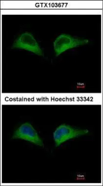
PSMD7 antibodyGTX103677
ApplicationsImmunoFluorescence, Western Blot, ImmunoCytoChemistry
ReactivityHuman
TargetPSMD7
- SizePrice
Product group Antibodies

PSMD7 / MOV34 AntibodyLS-C334006
ApplicationsImmunoFluorescence, Western Blot, ImmunoHistoChemistry
ReactivityHuman, Mouse
TargetPSMD7
- SizePrice

Product group Antibodies
Anti-PSMD7 AntibodyHPA049824
ApplicationsWestern Blot, ImmunoCytoChemistry, ImmunoHistoChemistry
ReactivityHuman
TargetPSMD7
- SizePrice
Product group Antibodies
PSMD7 antibodyGTX114902
ApplicationsWestern Blot
ReactivityHuman
TargetPSMD7
- SizePrice