

Psoriasin antibody, Internal
GTX89325
ApplicationsWestern Blot, ImmunoHistoChemistry, ImmunoHistoChemistry Paraffin
Product group Antibodies
ReactivityHuman
TargetS100A7
Overview
- SupplierGeneTex
- Product NamePsoriasin antibody, Internal
- Delivery Days Customer9
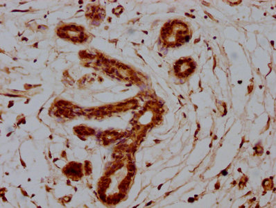
- Application Supplier NoteWB: 0.3-1microg/ml. IHC-P: 3.75microg/ml. *Optimal dilutions/concentrations should be determined by the researcher.Not tested in other applications.
- ApplicationsWestern Blot, ImmunoHistoChemistry, ImmunoHistoChemistry Paraffin
- CertificationResearch Use Only
- ClonalityPolyclonal
- Concentration0.50 mg/ml
- ConjugateUnconjugated
- Gene ID6278
- Target nameS100A7
- Target descriptionS100 calcium binding protein A7
- Target synonymsPSOR1, S100A7c, protein S100-A7, psoriasin 1
- HostGoat
- IsotypeIgG
- Protein IDP31151
- Protein NameProtein S100-A7
- Scientific DescriptionThe protein encoded by this gene is a member of the S100 family of proteins containing 2 EF-hand calcium-binding motifs. S100 proteins are localized in the cytoplasm and/or nucleus of a wide range of cells, and involved in the regulation of a number of cellular processes such as cell cycle progression and differentiation. S100 genes include at least 13 members which are located as a cluster on chromosome 1q21. This protein differs from the other S100 proteins of known structure in its lack of calcium binding ability in one EF-hand at the N-terminus. The protein is overexpressed in hyperproliferative skin diseases, exhibits antimicrobial activities against bacteria and induces immunomodulatory activities. [provided by RefSeq, Nov 2014]
- ReactivityHuman
- Storage Instruction-20°C or -80°C,2°C to 8°C
- UNSPSC12352203
Datasheet
Related products
Product group Antibodies
Anti-S100A7 Antibody144-65426
ApplicationsImmunoHistoChemistry
ReactivityHuman, Mouse, Rat
TargetS100A7
- SizePrice
Product group Antibodies
S100A7 Polyclonal AntibodyCAC11694
ApplicationsImmunoFluorescence, ELISA, ImmunoHistoChemistry
TargetS100A7
- SizePrice
Product group Antibodies
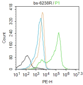
S100A7 Polyclonal AntibodyBS-6238R
ApplicationsFlow Cytometry, ImmunoFluorescence, ELISA, ImmunoCytoChemistry, ImmunoHistoChemistry, ImmunoHistoChemistry Frozen, ImmunoHistoChemistry Paraffin
ReactivityBovine, Equine, Human
TargetS100A7
- SizePrice
Product group Antibodies
S100A7 AntibodyCSB-PA020635HA01HU
ApplicationsImmunoFluorescence, ELISA, ImmunoHistoChemistry
ReactivityHuman
TargetS100A7
- SizePrice
![WB analysis of (A) MCF-10a and (B) MCF-7 cell lysate using GTX13680 Psoriasin antibody [47C1068]. Dilution : 1 μg/ml](https://www.genetex.com/upload/website/prouct_img/normal/GTX13680/GTX13680_975_WB_w_23060620_152.webp)
Product group Antibodies
Psoriasin antibody [47C1068]GTX13680
ApplicationsFlow Cytometry, ImmunoFluorescence, ImmunoPrecipitation, Western Blot, ImmunoCytoChemistry, ImmunoHistoChemistry, ImmunoHistoChemistry Frozen, ImmunoHistoChemistry Paraffin
ReactivityHuman
TargetS100A7
- SizePrice
Product group Antibodies

S100A7 / Psoriasin AntibodyLS-C831555
ApplicationsImmunoHistoChemistry
ReactivityHuman
TargetS100A7
- SizePrice

Product group Antibodies
Anti-S100A7 AntibodyHPA006997
ApplicationsImmunoHistoChemistry
ReactivityHuman
TargetS100A7
- SizePrice

Product group Antibodies
Anti-Psoriasin/S100A7 Antibody Picoband(r)A02369-1-CARRIER-FREE
ApplicationsFlow Cytometry, ImmunoFluorescence, Western Blot, ELISA, ImmunoCytoChemistry, ImmunoHistoChemistry
ReactivityHuman, Mouse, Rat
TargetS100A7
- SizePrice