

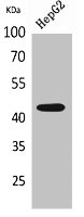
Western Blot analysis of HepG2 cells using EP1 Polyclonal Antibody.
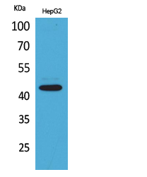
Western Blot analysis of HepG2 cells using EP1 Polyclonal Antibody.
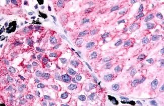
PTGER1 Antibody
CSB-PA005788
ApplicationsWestern Blot, ELISA
Product group Antibodies
ReactivityHuman
TargetPTGER1
Overview
- SupplierCusabio
- Product NamePTGER1 Antibody
- Delivery Days Customer20
- ApplicationsWestern Blot, ELISA
- CertificationResearch Use Only
- ClonalityPolyclonal
- ConjugateUnconjugated
- Gene ID5731
- Target namePTGER1
- Target descriptionprostaglandin E receptor 1
- Target synonymsEP1, prostaglandin E2 receptor EP1 subtype, PGE receptor, EP1 subtype, PGE2 receptor EP1 subtype, prostaglandin E receptor 1 (subtype EP1), 42kD, prostaglandin E receptor 1 (subtype EP1), 42kDa, prostaglandin E receptor 1, subtype EP1, prostanoid EP1 receptor
- HostRabbit
- IsotypeIgG
- Protein IDP34995
- Protein NameProstaglandin E2 receptor EP1 subtype
- ReactivityHuman
- Storage Instruction-20°C or -80°C
- UNSPSC41116161
Related products
Product group Antibodies
Anti-PTGER1 AntibodyA100335
ApplicationsWestern Blot, ELISA
ReactivityHuman
- SizePrice
Product group Antibodies

ApplicationsELISA, ImmunoHistoChemistry
ReactivityHuman
TargetPTGER1
- SizePrice
Product group Antibodies

PTGER1 / EP1 AntibodyLS-C405554
ApplicationsWestern Blot, ELISA
ReactivityHuman, Mouse, Rat
TargetPTGER1
- SizePrice
Product group Antibodies
References
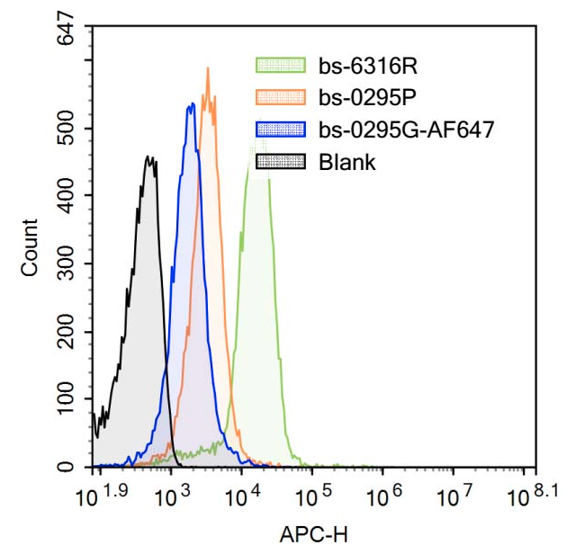
PTGER1 Polyclonal AntibodyBS-6316R
ApplicationsFlow Cytometry, ImmunoFluorescence, Western Blot, ELISA, ImmunoCytoChemistry, ImmunoHistoChemistry, ImmunoHistoChemistry Frozen, ImmunoHistoChemistry Paraffin
ReactivityCanine, Human, Mouse, Rat
TargetPTGER1
- SizePrice
Product group Antibodies
Anti-Prostaglandin E Receptor EP1/PTGER1 Antibody Picoband(r)PA1583-CARRIER-FREE
ApplicationsWestern Blot
ReactivityHamster, Human
TargetPTGER1
- SizePrice
Product group Antibodies
ApplicationsImmunoHistoChemistry, ImmunoHistoChemistry Paraffin
ReactivityHuman
TargetPTGER1
- SizePrice
Product group Antibodies
Anti-Rat PTGER1 Antibody144-12310
ApplicationsWestern Blot
ReactivityHuman, Mouse, Rat
TargetPTGER1
- SizePrice