

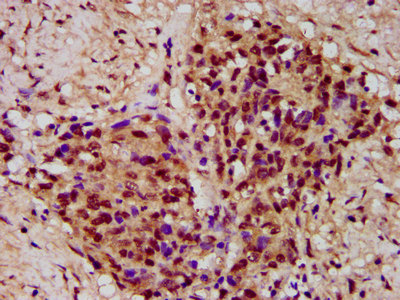
IHC image of CSB-PA883586LA01HU diluted at 1:300 and staining in paraffin-embedded human ovarian cancer performed on a Leica BondTM system. After dewaxing and hydration, antigen retrieval was mediated by high pressure in a citrate buffer (pH 6.0). Section was blocked with 10% normal goat serum 30min at RT. Then primary antibody (1% BSA) was incubated at 4°C overnight. The primary is detected by a biotinylated secondary antibody and visualized using an HRP conjugated SP system.
. Section was blocked with 10% normal goat serum 30min at RT. Then primary antibody (1% BSA) was incubated at 4°C overnight. The primary is detected by a biotinylated secondary antibody and visualized using an HRP conjugated SP system.")
.")
IHC image of CSB-PA883586LA01HU diluted at 1:300 and staining in paraffin-embedded human ovarian cancer performed on a Leica BondTM system. After dewaxing and hydration, antigen retrieval was mediated by high pressure in a citrate buffer (pH 6.0). Section was blocked with 10% normal goat serum 30min at RT. Then primary antibody (1% BSA) was incubated at 4°C overnight. The primary is detected by a biotinylated secondary antibody and visualized using an HRP conjugated SP system.
PYGO2 Antibody
CSB-PA883586LA01HU
ApplicationsImmunoFluorescence, ELISA, ImmunoHistoChemistry
Product group Antibodies
ReactivityHuman
TargetPYGO2
Overview
- SupplierCusabio
- Product NamePYGO2 Antibody
- Delivery Days Customer20
- ApplicationsImmunoFluorescence, ELISA, ImmunoHistoChemistry
- CertificationResearch Use Only
- ClonalityPolyclonal
- ConjugateUnconjugated
- Gene ID90780
- Target namePYGO2
- Target descriptionpygopus family PHD finger 2
- Target synonyms1190004M21Rik, pygopus homolog 2, pygopus 2
- HostRabbit
- IsotypeIgG
- Protein IDQ9BRQ0
- Protein NamePygopus homolog 2
- Scientific DescriptionInvolved in signal transduction through the Wnt pathway.
- ReactivityHuman
- Storage Instruction-20°C or -80°C
- UNSPSC41116161
Related products

Product group Antibodies
ApplicationsImmunoFluorescence, Western Blot, ImmunoCytoChemistry, ImmunoHistoChemistry
ReactivityHuman, Mouse
TargetPYGO2
- SizePrice

Product group Antibodies
Anti-PYGO2 AntibodyHPA023689
ApplicationsImmunoCytoChemistry, ImmunoHistoChemistry
ReactivityHuman
TargetPYGO2
- SizePrice
Product group Antibodies

PYGO2 / Pygopus 2 Antibody (HRP)LS-C673144
ReactivityHuman
TargetPYGO2
- SizePrice
Product group Antibodies
ApplicationsImmunoPrecipitation, Western Blot, ImmunoCytoChemistry, ImmunoHistoChemistry
TargetPYGO2
- SizePrice
Product group Antibodies
PYGO2 AntibodyPACO58572
ApplicationsImmunoFluorescence, ELISA, ImmunoHistoChemistry
ReactivityHuman
TargetPYGO2
- SizePrice
Product group Antibodies
Pygopus 2 Recombinant AntibodyBSM-61113R
ApplicationsFlow Cytometry, ImmunoFluorescence, Western Blot, ImmunoCytoChemistry, ImmunoHistoChemistry, ImmunoHistoChemistry Frozen, ImmunoHistoChemistry Paraffin
TargetPYGO2
- SizePrice
Product group Antibodies
Pygopus 2 antibodyGTX116847
ApplicationsImmunoFluorescence, Western Blot, ImmunoCytoChemistry
ReactivityHuman
TargetPYGO2
- SizePrice