![QKI antibody [GT1589] detects QKI protein at cytoplasm and nucleus in mouse brain by immunohistochemical analysis. Sample: Paraffin-embedded mouse brain. QKI antibody [GT1589] (GTX633920) diluted at 1:200.
Antigen Retrieval: Citrate buffer, pH 6.0, 15 min QKI antibody [GT1589] detects QKI protein at cytoplasm and nucleus in mouse brain by immunohistochemical analysis. Sample: Paraffin-embedded mouse brain. QKI antibody [GT1589] (GTX633920) diluted at 1:200.
Antigen Retrieval: Citrate buffer, pH 6.0, 15 min](https://www.genetex.com/upload/website/prouct_img/normal/GTX633920/GTX633920_42709_20170405_IHC-P_M_w_23061202_160.webp)
QKI antibody [GT1589] detects QKI protein at cytoplasm and nucleus in mouse brain by immunohistochemical analysis. Sample: Paraffin-embedded mouse brain. QKI antibody [GT1589] (GTX633920) diluted at 1:200.
Antigen Retrieval: Citrate buffer, pH 6.0, 15 min
QKI antibody [GT1589]
GTX633920
ApplicationsImmunoFluorescence, Western Blot, ImmunoCytoChemistry, ImmunoHistoChemistry, ImmunoHistoChemistry Frozen, ImmunoHistoChemistry Paraffin
Product group Antibodies
ReactivityHuman, Mouse, Rat
TargetQKI
Overview
- SupplierGeneTex
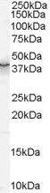
- Product NameQKI antibody [GT1589]
- Delivery Days Customer9
- Application Supplier NoteWB: 1:1000-1:10000. ICC/IF: 1:100-1:1000. IHC-P: 1:100-1:1000. IHC-Fr: 1:100-1:1000. *Optimal dilutions/concentrations should be determined by the researcher.Not tested in other applications.
- ApplicationsImmunoFluorescence, Western Blot, ImmunoCytoChemistry, ImmunoHistoChemistry, ImmunoHistoChemistry Frozen, ImmunoHistoChemistry Paraffin
- CertificationResearch Use Only
- ClonalityMonoclonal
- Clone IDGT1589
- Concentration1 mg/ml
- ConjugateUnconjugated
- Gene ID9444
- Target nameQKI
- Target descriptionQKI, KH domain containing RNA binding
- Target synonymsHqk, QK, QK1, QK3, hqkI, KH domain-containing RNA-binding protein QKI, QKI/LOC100132735 fusion, RNA binding protein HQK, homolog of mouse quaking QKI (KH domain RNA binding protein), quaking homolog, KH domain RNA binding
- HostMouse
- IsotypeIgG1
- Protein IDQ96PU8
- Protein NameKH domain-containing RNA-binding protein QKI
- Scientific DescriptionQKI belongs to a family of RNA-binding proteins that have an HNRNPK (MIM 600712) homology (KH) domain embedded in a 200-amino acid region called the GSG domain. Other members of this family include SAM68 (KHDRBS1; MIM 602489) and SF1 (MIM 601516) (Chen and Richard, 1998 [PubMed 9671495]). QKI proteins regulate RNA splicing, export of target RNAs from the nucleus, translation of proteins, and RNA stability (Lauriat et al., 2008 [PubMed 17918747]).[supplied by OMIM]
- ReactivityHuman, Mouse, Rat
- Storage Instruction-20°C or -80°C,2°C to 8°C
- UNSPSC41116161
![Whole cell extract (30 μg) was separated by 10% SDS-PAGE, and the membrane was blotted with QKI antibody [GT1589] (GTX633920) diluted at 1:3000. Whole cell extract (30 μg) was separated by 10% SDS-PAGE, and the membrane was blotted with QKI antibody [GT1589] (GTX633920) diluted at 1:3000.](https://www.genetex.com/upload/website/prouct_img/normal/GTX633920/GTX633920_42709_20170119_WB_w_23061202_582.webp)
![QKI antibody [GT1589] detects QKI protein expression by immunohistochemical analysis. Sample: Frozen-sectioned adult mouse cerebellum. Green: QKI protein stained by QKI antibody [GT1589] (GTX633920) diluted at 1:250. Red: Calbindin, stained by Calbindin antibody (GTX130856) diluted at 1:500. Blue: Fluoroshield with DAPI (GTX30920). QKI antibody [GT1589] detects QKI protein expression by immunohistochemical analysis. Sample: Frozen-sectioned adult mouse cerebellum. Green: QKI protein stained by QKI antibody [GT1589] (GTX633920) diluted at 1:250. Red: Calbindin, stained by Calbindin antibody (GTX130856) diluted at 1:500. Blue: Fluoroshield with DAPI (GTX30920).](https://www.genetex.com/upload/website/prouct_img/normal/GTX633920/GTX633920_42709_20170807_IHC-Fr_M_w_23061202_157.webp)
![QKI antibody [GT1589] detects QKI protein at cytoplasm and nucleus by immunofluorescent analysis. Sample: SK-N-AS cells were fixed in 4% paraformaldehyde at RT for 15 min. Green: QKI protein stained by QKI antibody [GT1589] (GTX633920) diluted at 1:500. Blue: Hoechst 33342 staining. Scale bar = 10 μm. QKI antibody [GT1589] detects QKI protein at cytoplasm and nucleus by immunofluorescent analysis. Sample: SK-N-AS cells were fixed in 4% paraformaldehyde at RT for 15 min. Green: QKI protein stained by QKI antibody [GT1589] (GTX633920) diluted at 1:500. Blue: Hoechst 33342 staining. Scale bar = 10 μm.](https://www.genetex.com/upload/website/prouct_img/normal/GTX633920/GTX633920_42709_20170412_IFA_w_23061202_645.webp)
![QKI antibody [GT1589] detects QKI protein at cytoplasm and nucleus in rat brain by immunohistochemical analysis. Sample: Paraffin-embedded rat brain. QKI antibody [GT1589] (GTX633920) diluted at 1:200.
Antigen Retrieval: Citrate buffer, pH 6.0, 15 min QKI antibody [GT1589] detects QKI protein at cytoplasm and nucleus in rat brain by immunohistochemical analysis. Sample: Paraffin-embedded rat brain. QKI antibody [GT1589] (GTX633920) diluted at 1:200.
Antigen Retrieval: Citrate buffer, pH 6.0, 15 min](https://www.genetex.com/upload/website/prouct_img/normal/GTX633920/GTX633920_42709_20170405_IHC-P_R_w_23061202_639.webp)
![Non-transfected (–) and transfected (+) 293T whole cell extracts (30 μg) were separated by 10% SDS-PAGE, and the membrane was blotted with QKI antibody [GT1589] (GTX633920) diluted at 1:5000. The HRP-conjugated anti-mouset IgG antibody (GTX213111-01) was used to detect the primary antibody. Non-transfected (–) and transfected (+) 293T whole cell extracts (30 μg) were separated by 10% SDS-PAGE, and the membrane was blotted with QKI antibody [GT1589] (GTX633920) diluted at 1:5000. The HRP-conjugated anti-mouset IgG antibody (GTX213111-01) was used to detect the primary antibody.](https://www.genetex.com/upload/website/prouct_img/normal/GTX633920/GTX633920_42709_20180817_WB_shRNA_watermark_w_23061202_323.webp)


![QKI antibody [N2C3] detects QKI protein at cytoplasm and nucleus by immunofluorescent analysis. Sample: SK-N-SH cells were fixed in 4% paraformaldehyde at RT for 15 min. Green: QKI protein stained by QKI antibody [N2C3] (GTX115969) diluted at 1:500. Blue: Hoechst 33342 staining. Scale bar = 10 μm.](https://www.genetex.com/upload/website/prouct_img/normal/GTX115969/GTX115969_40289_20161130_IFA_w_23060519_615.webp)