

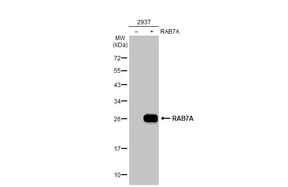
Non-transfected (–) and transfected (+) 293T whole cell extracts (30 μg) were separated by 12% SDS-PAGE, and the membrane was blotted with RAB7A antibody (GTX132548) diluted at 1:5000. The HRP-conjugated anti-rabbit IgG antibody (GTX213110-01) was used to detect the primary antibody.
 diluted at 1:500.
Antigen Retrieval: Citrate buffer, pH 6.0, 15 min")
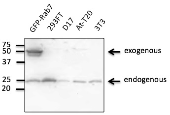
 were separated by 12% SDS-PAGE, and the membrane was blotted with Rab7 antibody (GTX132548) diluted at 1:1000.")
 diluted at 1:500.
Antigen Retrieval: Citrate buffer, pH 6.0, 15 min")
![RAB7A antibody detects RAB7A protein by immunofluorescent analysis. Sample: DIV9 rat E18 primary cortical neuron cells were fixed in 4% paraformaldehyde at RT for 15 min. Green: RAB7A stained by RAB7A antibody (GTX132548) diluted at 1:500. Red: beta Tubulin 3/ Tuj1, stained by beta Tubulin 3/ Tuj1 antibody [GT1338] (GTX631831) diluted at 1:500. Blue: Fluoroshield with DAPI (GTX30920).](https://www.genetex.com/upload/website/prouct_img/normal/GTX132548/GTX132548_42452_20171115_ICC_IF_R_w_23060523_949.webp "RAB7A antibody detects RAB7A protein by immunofluorescent analysis. Sample: DIV9 rat E18 primary cortical neuron cells were fixed in 4% paraformaldehyde at RT for 15 min. Green: RAB7A stained by RAB7A antibody (GTX132548) diluted at 1:500. Red: beta Tubulin 3/ Tuj1, stained by beta Tubulin 3/ Tuj1 antibody [GT1338] (GTX631831) diluted at 1:500. Blue: Fluoroshield with DAPI (GTX30920).")
 and transfected (+) 293T whole cell extracts (50 μg) were separated by 12% SDS-PAGE, and the membrane was blotted with RAB7A antibody (GTX132548) diluted at 1:1000. The HRP-conjugated anti-rabbit IgG antibody (GTX213110-01) was used to detect the primary antibody, and the signal was developed with Trident ECL plus-Enhanced.")
 were separated by 12% SDS-PAGE, and the membrane was blotted with RAB7A antibody (GTX132548) diluted at 1:1000 and competitor's antibody (SC-10767) diluted by 1:200.")
 diluted at 1:500. Blue: Hoechst 33342 staining.")
Non-transfected (–) and transfected (+) 293T whole cell extracts (30 μg) were separated by 12% SDS-PAGE, and the membrane was blotted with RAB7A antibody (GTX132548) diluted at 1:5000. The HRP-conjugated anti-rabbit IgG antibody (GTX213110-01) was used to detect the primary antibody.
RAB7A antibody
GTX132548
ApplicationsImmunoFluorescence, Western Blot, ImmunoCytoChemistry, ImmunoHistoChemistry, ImmunoHistoChemistry Paraffin
Product group Antibodies
ReactivityHuman, Mouse, Rat
TargetRAB7A
Overview
- SupplierGeneTex
- Product NameRAB7A antibody
- Delivery Days Customer9
- Application Supplier NoteWB: 1:500-1:3000. ICC/IF: 1:100-1:1000. IHC-P: 1:100-1:1000. *Optimal dilutions/concentrations should be determined by the researcher.Not tested in other applications.
- ApplicationsImmunoFluorescence, Western Blot, ImmunoCytoChemistry, ImmunoHistoChemistry, ImmunoHistoChemistry Paraffin
- CertificationResearch Use Only
- ClonalityPolyclonal
- Concentration0.36 mg/ml
- ConjugateUnconjugated
- Gene ID7879
- Target nameRAB7A
- Target descriptionRAB7A, member RAS oncogene family
- Target synonymsCMT2B, PRO2706, RAB7, ras-related protein Rab-7a, RAB7, member RAS oncogene family, Ras-associated protein RAB7
- HostRabbit
- IsotypeIgG
- Protein IDP51149
- Protein NameRas-related protein Rab-7a
- Scientific DescriptionRAB family members are small, RAS-related GTP-binding proteins that are important regulators of vesicular transport. Each RAB protein targets multiple proteins that act in exocytic / endocytic pathways. This gene encodes a RAB family member that regulates vesicle traffic in the late endosomes and also from late endosomes to lysosomes. This encoded protein is also involved in the cellular vacuolation of the VacA cytotoxin of Helicobacter pylori. Mutations at highly conserved amino acid residues in this gene have caused some forms of Charcot-Marie-Tooth (CMT) type 2 neuropathies. [provided by RefSeq, Jul 2008]
- ReactivityHuman, Mouse, Rat
- Storage Instruction-20°C or -80°C,2°C to 8°C
- UNSPSC12352203
Datasheet
Related products
Product group Antibodies
Anti-RAB7A Antibody144-60102
ApplicationsImmunoFluorescence, Western Blot, ImmunoHistoChemistry
ReactivityHuman, Mouse, Rat
TargetRAB7A
- SizePrice

Product group Antibodies
Anti-RAB7/RAB7A Antibody Picoband(r)A02409-1-CARRIER-FREE
ApplicationsFlow Cytometry, Western Blot, ELISA, ImmunoHistoChemistry
ReactivityHuman, Mouse, Rat
TargetRAB7A
- SizePrice
Product group Antibodies
RAB7A antibodyGTX130847
ApplicationsWestern Blot, ImmunoHistoChemistry, ImmunoHistoChemistry Paraffin
ReactivityHuman, Mouse
TargetRAB7A
- SizePrice
Product group Antibodies
RAB7A antibody [AT10E4]GTX57709
ApplicationsFlow Cytometry, ImmunoFluorescence, Western Blot, ImmunoCytoChemistry
ReactivityHuman
TargetRAB7A
- SizePrice
Product group Antibodies
RAB7A Polyclonal AntibodyBS-6703R
ApplicationsImmunoFluorescence, Western Blot, ELISA, ImmunoCytoChemistry
ReactivityHuman, Mouse, Rat
TargetRAB7A
- SizePrice
Product group Antibodies
Anti-RAB7A AntibodyA121566
ApplicationsImmunoFluorescence, Western Blot
ReactivityCanine, Human, Monkey, Mouse, Rat
- SizePrice
Product group Antibodies

RAB7A / RAB7 AntibodyLS-C747860
ApplicationsWestern Blot
ReactivityHuman, Mouse, Rat
TargetRAB7A
- SizePrice
Product group Antibodies
RAB7A AntibodyCSB-PA019219LA01HU
ApplicationsImmunoFluorescence, ELISA, ImmunoHistoChemistry
ReactivityHuman
TargetRAB7A
- SizePrice