

Rabbit anti Human Sphingosine 1 Phosphate Receptor 1 (EDG-1) CT
X1093P
ApplicationsWestern Blot
Product group Antibodies
ReactivityHuman, Mouse, Rat
TargetS1PR1
Product X1093P is not available
Product not available
There may be an alternative product available, please contact our technical support team.
Overview
- SupplierNordic-MUbio
- Product NameRabbit anti Human Sphingosine 1 Phosphate Receptor 1 (EDG-1) CT
- Delivery Days Customer7
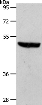
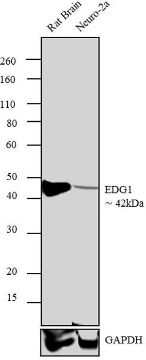
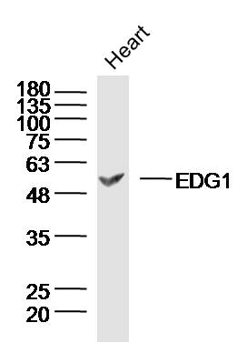
- Application Supplier NoteDetects EDG1 receptors at concentration of 5-10 microg/ml by Western blot using a human EDG1 receptor transfected cell line. Detects an approximately 44 kDa band in RH7777 cells transfected with full length human EDG1. Due to low expression of EDG receptors, we recommend use of Pierce Femto Signal substrate for western blot development. Optimal concentration should be evaluated by serial dilutions.
- ApplicationsWestern Blot
- Applications SupplierWestern Blotting;Western Blotting
- CertificationResearch Use Only
- ClonalityPolyclonal
- ConjugateUnconjugated
- Gene ID1901
- Target nameS1PR1
- Target descriptionsphingosine-1-phosphate receptor 1
- Target synonymsCD363, CHEDG1, D1S3362, ECGF1, EDG-1, EDG1, S1P1, sphingosine 1-phosphate receptor 1, S1P receptor Edg-1, endothelial differentiation G-protein coupled receptor 1, endothelial differentiation, sphingolipid G-protein-coupled receptor, 1, sphingosine 1-phosphate receptor EDG1, sphingosine 1-phosphate receptor Edg-1
- HostRabbit
- IsotypeIgG
- Protein IDP21453
- Protein NameSphingosine 1-phosphate receptor 1
- Scientific DescriptionEndothelial cell differentiation gene 1 C-terminal
- Shelf life instructionSee expiration date on vial
- ReactivityHuman, Mouse, Rat
- Reactivity SupplierHuman;Mouse;Rat
- Reactivity Supplier NoteSynthetic peptide derived from the C-terminal of the EDG-1 receptor
- UNSPSC12352203
Related products
Product group Antibodies
Anti-S1PR1 Antibody144-12935
ApplicationsWestern Blot
ReactivityHuman, Mouse
TargetS1PR1
- SizePrice
Product group Antibodies
S1PR1 Polyclonal AntibodyCAC13921
ApplicationsImmunoFluorescence, Western Blot, ELISA, ImmunoHistoChemistry
ReactivityMouse
TargetS1PR1
- SizePrice

Product group Antibodies
References
ApplicationsWestern Blot
ReactivityHuman, Mouse, Rat
TargetS1PR1
- SizePrice
Product group Antibodies
Anti-S1PR1 AntibodyA43229
ApplicationsWestern Blot
ReactivityHuman, Mouse
- SizePrice

Product group Antibodies
S1PR1 AntibodyCSB-PA002247
ApplicationsImmunoFluorescence, Western Blot, ELISA
ReactivityHuman, Mouse, Rat
TargetS1PR1
- SizePrice
Product group Antibodies
EDG1 antibodyGTX11424
ApplicationsFlow Cytometry, ImmunoFluorescence, ImmunoPrecipitation, Western Blot, ImmunoCytoChemistry, ImmunoHistoChemistry, ImmunoHistoChemistry Frozen
ReactivityHuman, Mouse, Rat
TargetS1PR1
- SizePrice

Product group Antibodies
Anti-S1PR1 AntibodyHPA075568
ApplicationsImmunoCytoChemistry
ReactivityHuman
TargetS1PR1
- SizePrice
Product group Antibodies
References
EDG1 Polyclonal AntibodyBS-7112R
ApplicationsImmunoFluorescence, Western Blot, ELISA, ImmunoCytoChemistry, ImmunoHistoChemistry, ImmunoHistoChemistry Frozen, ImmunoHistoChemistry Paraffin
ReactivityCanine, Equine, Human, Mouse, Rat
TargetS1PR1
- SizePrice