

Rabbit anti-TAK1 Antibody, Affinity Purified
A301-915A
ApplicationsImmunoPrecipitation
Product group Antibodies
ReactivityBovine, Human, Mouse, Primate, Rat
TargetMAP3K7
Product A301-915A is not available
Product not available
There may be an alternative product available, please contact our technical support team.
Overview
- SupplierBethyl Laboratories
- Product NameRabbit anti-TAK1 Antibody, Affinity Purified
- Delivery Days Customer9
- ApplicationsImmunoPrecipitation
- CertificationResearch Use Only
- ClonalityPolyclonal
- Concentration1000 ug/ml
- ConjugateUnconjugated
- Estimated Purity...
- Gene ID6885
- Target nameMAP3K7
- Target descriptionmitogen-activated protein kinase kinase kinase 7
- Target synonymsCSCF, FMD2, MEKK7, TAK1, TGF1a, mitogen-activated protein kinase kinase kinase 7, TGF-beta activated kinase 1, transforming growth factor-beta-activated kinase 1
- HostRabbit
- IsotypeIgG
- Protein IDO43318
- Protein NameMitogen-activated protein kinase kinase kinase 7
- ReactivityBovine, Human, Mouse, Primate, Rat
- Storage Instruction2°C to 8°C
- UNSPSC12352203
Related products
Product group Antibodies
ApplicationsWestern Blot, ELISA, ImmunoHistoChemistry
TargetMAP3K7
- SizePrice
Product group Antibodies
References
ApplicationsFlow Cytometry, ImmunoFluorescence, Western Blot, ELISA, ImmunoCytoChemistry, ImmunoHistoChemistry, ImmunoHistoChemistry Frozen, ImmunoHistoChemistry Paraffin
ReactivityBovine, Chicken, Equine, Human, Mouse, Porcine, Rabbit, Rat
TargetMAP3K7
- SizePrice
Product group Antibodies
Anti-MAP3K7 Antibody144-62173
ApplicationsWestern Blot
ReactivityHuman, Mouse
TargetMAP3K7
- SizePrice
Product group Antibodies

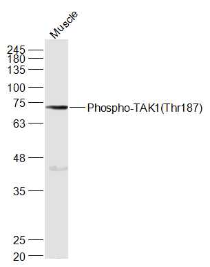
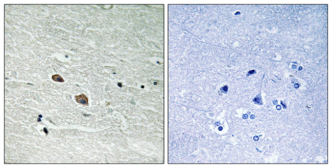
MAP3K7 (Phospho-Thr187) AntibodyABX012607
ApplicationsWestern Blot, ELISA
- SizePrice
Product group Antibodies
Anti-MAP3K7 AntibodyA95622
ApplicationsWestern Blot, ELISA, ImmunoHistoChemistry
ReactivityHuman, Mouse, Rat
- SizePrice
Product group Antibodies

ApplicationsFlow Cytometry, Western Blot, ELISA
ReactivityBovine, Human, Mouse, Rat
TargetMAP3K7
- SizePrice
Product group Antibodies
References
TAK1 antibodyGTX107452
ApplicationsWestern Blot
ReactivityHuman
TargetMAP3K7
- SizePrice
Product group Antibodies

MAP3K7 / TAK1 AntibodyLS-C772775
ApplicationsWestern Blot, ELISA, ImmunoHistoChemistry, ImmunoHistoChemistry Paraffin
ReactivityHuman, Mouse, Rat
TargetMAP3K7
- SizePrice