

IHC image of CSB-PA009605LA01HU diluted at 1:500 and staining in paraffin-embedded human small intestine tissue performed on a Leica BondTM system. After dewaxing and hydration, antigen retrieval was mediated by high pressure in a citrate buffer (pH 6.0). Section was blocked with 10% normal goat serum 30min at RT. Then primary antibody (1% BSA) was incubated at 4°C overnight. The primary is detected by a biotinylated secondary antibody and visualized using an HRP conjugated SP system.
.")
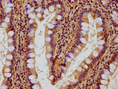
IHC image of CSB-PA009605LA01HU diluted at 1:500 and staining in paraffin-embedded human small intestine tissue performed on a Leica BondTM system. After dewaxing and hydration, antigen retrieval was mediated by high pressure in a citrate buffer (pH 6.0). Section was blocked with 10% normal goat serum 30min at RT. Then primary antibody (1% BSA) was incubated at 4°C overnight. The primary is detected by a biotinylated secondary antibody and visualized using an HRP conjugated SP system.
RACK1 Antibody
CSB-PA009605LA01HU
ApplicationsImmunoFluorescence, ELISA, ImmunoHistoChemistry
Product group Antibodies
ReactivityHuman
TargetRACK1
Overview
- SupplierCusabio
- Product NameRACK1 Antibody
- Delivery Days Customer20
- ApplicationsImmunoFluorescence, ELISA, ImmunoHistoChemistry
- CertificationResearch Use Only
- ClonalityPolyclonal
- ConjugateUnconjugated
- Gene ID10399
- Target nameRACK1
- Target descriptionreceptor for activated C kinase 1
- Target synonymsGNB2L1, Gnb2-rs1, H12.3, HLC-7, PIG21, small ribosomal subunit protein RACK1, cell proliferation-inducing gene 21 protein, guanine nucleotide binding protein (G protein), beta polypeptide 2-like 1, guanine nucleotide binding protein beta polypeptide 2-like 1, guanine nucleotide-binding protein subunit beta-2-like 1, guanine nucleotide-binding protein subunit beta-like protein 12.3, human lung cancer oncogene 7 protein, lung cancer oncogene 7, proliferation-inducing gene 21, protein homologous to chicken B complex protein, guanine nucleotide binding, receptor of activated protein C kinase 1, receptor of activated protein kinase C 1
- HostRabbit
- IsotypeIgG
- Protein IDP63244
- Protein NameSmall ribosomal subunit protein RACK1
- Scientific DescriptionInvolved in the recruitment, assembly and/or regulation of a variety of signaling molecules. Interacts with a wide variety of proteins and plays a role in many cellular processes. Component of the 40S ribosomal subunit involved in translational repression (PubMed:23636399). Involved in the initiation of the ribosome quality control (RQC), a pathway that takes place when a ribosome has stalled during translation, by promoting ubiquitination of a subset of 40S ribosomal subunits (PubMed:28132843). Binds to and stabilizes activated protein kinase C (PKC), increasing PKC-mediated phosphorylation. May recruit activated PKC to the ribosome, leading to phosphorylation of EIF6. Inhibits the activity of SRC kinases including SRC, LCK and YES1. Inhibits cell growth by prolonging the G0/G1 phase of the cell cycle. Enhances phosphorylation of BMAL1 by PRKCA and inhibits transcriptional activity of the BMAL1-CLOCK heterodimer. Facilitates ligand-independent nuclear translocation of AR following PKC activation, represses AR transactivation activity and is required for phosphorylation of AR by SRC. Modulates IGF1R-dependent integrin signaling and promotes cell spreading and contact with the extracellular matrix. Involved in PKC-dependent translocation of ADAM12 to the cell membrane. Promotes the ubiquitination and proteasome-mediated degradation of proteins such as CLEC1B and HIF1A. Required for VANGL2 membrane localization, inhibits Wnt signaling, and regulates cellular polarization and oriented cell division during gastrulation. Required for PTK2/FAK1 phosphorylation and dephosphorylation. Regulates internalization of the muscarinic receptor CHRM2. Promotes apoptosis by increasing oligomerization of BAX and disrupting the interaction of BAX with the anti-apoptotic factor BCL2L. Inhibits TRPM6 channel activity. Regulates cell surface expression of some GPCRs such as TBXA2R. Plays a role in regulation of FLT1-mediated cell migration. Involved in the transport of ABCB4 from the Golgi to the apical bile canalicular membrane (PubMed:19674157). Promotes migration of breast carcinoma cells by binding to and activating RHOA (PubMed:20499158).
- ReactivityHuman
- Storage Instruction-20°C or -80°C
- UNSPSC41116161
Related products
Product group Antibodies
Anti-GNB2L1 (N-term) Antibody102-27485
ApplicationsWestern Blot, ImmunoHistoChemistry, ImmunoHistoChemistry Paraffin
TargetRACK1
- SizePrice
Product group Antibodies
RACK1 Recombinant AntibodyBSM-61942R
ApplicationsFlow Cytometry, ImmunoFluorescence, ImmunoCytoChemistry, ImmunoHistoChemistry, ImmunoHistoChemistry Frozen, ImmunoHistoChemistry Paraffin
ReactivityHuman, Mouse, Rat
TargetRACK1
- SizePrice
Product group Antibodies

Goat anti-GNB2L1EB10690
ApplicationsWestern Blot, ELISA, ImmunoHistoChemistry
ReactivityBovine, Canine, Human, Mouse, Rat
TargetRACK1
- SizePrice
Product group Antibodies
RACK1 antibodyGTX102160
ApplicationsWestern Blot
ReactivityHuman
TargetRACK1
- SizePrice

Product group Antibodies
Anti-RACK1-25ulHPA021676
ApplicationsWestern Blot, ImmunoCytoChemistry
ReactivityHuman, Mouse
- SizePrice

Product group Antibodies
Anti-RACK1 GNB2L1 Antibody Picoband(r)PB9243-CARRIER-FREE
ApplicationsFlow Cytometry, ImmunoFluorescence, Western Blot, ImmunoCytoChemistry
ReactivityBovine, Canine, Chicken, Equine, Human, Monkey, Mouse, Rabbit, Rat
TargetRACK1
- SizePrice
Product group Antibodies

GNB2L1 / RACK1 AntibodyLS-C746714
ApplicationsImmunoFluorescence, Western Blot, ImmunoHistoChemistry
ReactivityHuman, Mouse
TargetRACK1
- SizePrice
Product group Antibodies
ApplicationsWestern Blot, ELISA
ReactivityHuman
TargetRACK1
- SizePrice