


Gel: 12%SDS-PAGE Lysate: 50 microg Lane 1-2: NIH/3T3 cells Jurkat cells Primary antibody: TA364954 (RACK1 Antibody) at dilution 1/300 Secondary antibody: Goat anti rabbit IgG at 1/8000 dilution Exposure time: 20 seconds
Gel: 12%SDS-PAGE Lysate: 50 microg Lane 1-2: NIH/3T3 cells Jurkat cells Primary antibody: TA364954 (RACK1 Antibody) at dilution 1/300 Secondary antibody: Goat anti rabbit IgG at 1/8000 dilution Exposure time: 20 seconds
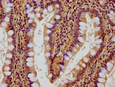
RACK1 Rabbit Polyclonal Antibody
TA364954
Overview
- SupplierOriGene
- Product NameRACK1 Rabbit Polyclonal Antibody
- Delivery Days Customer14
- ApplicationsWestern Blot
- CertificationResearch Use Only
- ClonalityPolyclonal
- Gene ID10399
- Target nameRACK1
- Target descriptionreceptor for activated C kinase 1
- Target synonymscell proliferation-inducing gene 21 protein; GNB2L1; Gnb2-rs1; guanine nucleotide binding protein (G protein), beta polypeptide 2-like 1; guanine nucleotide binding protein beta polypeptide 2-like 1; guanine nucleotide-binding protein subunit beta-2-like 1; guanine nucleotide-binding protein subunit beta-like protein 12.3; H12.3; HLC-7; human lung cancer oncogene 7 protein; lung cancer oncogene 7; PIG21; proliferation-inducing gene 21; protein homologous to chicken B complex protein, guanine nucleotide binding; receptor of activated protein C kinase 1; receptor of activated protein kinase C 1; small ribosomal subunit protein RACK1
- HostRabbit
- IsotypeIgG
- Protein IDP63244
- Protein NameReceptor of activated protein C kinase 1
- Scientific DescriptionRACK1 rabbit polyclonal antibody
- Storage Instruction-20°C
- UNSPSC12352203
Related products
Product group Antibodies
RACK1 AntibodyCSB-PA009605LA01HU
ApplicationsImmunoFluorescence, ELISA, ImmunoHistoChemistry
ReactivityHuman
TargetRACK1
- SizePrice
Product group Antibodies
Anti-GNB2L1 (N-term) Antibody102-27485
ApplicationsWestern Blot, ImmunoHistoChemistry, ImmunoHistoChemistry Paraffin
TargetRACK1
- SizePrice
Product group Antibodies
RACK1 Recombinant AntibodyBSM-61942R
ApplicationsFlow Cytometry, ImmunoFluorescence, Western Blot, ImmunoHistoChemistry, ImmunoHistoChemistry Frozen, ImmunoHistoChemistry Paraffin
TargetRACK1
- SizePrice

Product group Antibodies
Anti-RACK1 AntibodyHPA021676
ApplicationsWestern Blot, ImmunoCytoChemistry
ReactivityHuman, Mouse
TargetRACK1
- SizePrice

Product group Antibodies
Anti-RACK1 GNB2L1 Antibody Picoband(r)PB9243-CARRIER-FREE
ApplicationsFlow Cytometry, ImmunoFluorescence, Western Blot, ImmunoCytoChemistry
TargetRACK1
- SizePrice
Product group Antibodies

Goat anti-GNB2L1EB10690
ApplicationsWestern Blot, ELISA, ImmunoHistoChemistry
TargetRACK1
- SizePrice
Product group Antibodies
References
RACK1 antibodyGTX102160
ApplicationsWestern Blot
TargetRACK1
- SizePrice
Product group Antibodies
Anti-RACK1 AntibodyA283476
ApplicationsWestern Blot, ImmunoHistoChemistry, ImmunoHistoChemistry Paraffin
- SizePrice
Product group Antibodies

GNB2L1 / RACK1 AntibodyLS-C746714
ApplicationsImmunoFluorescence, Western Blot, ImmunoHistoChemistry
TargetRACK1
- SizePrice