

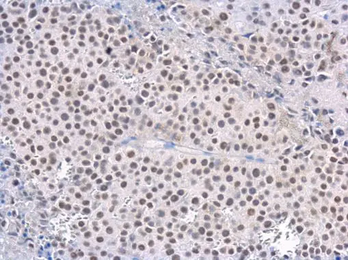
Rad50 antibody [13B3] detects Rad50 protein at nucleus in CAL 27 xenograft by immunohistochemical analysis. Sample: Paraffin-embedded CAL 27 xenograft. Rad50 antibody [13B3] (GTX70228) diluted at 1:200.
Antigen Retrieval: Citrate buffer, pH 6.0, 15 min
![Rad50 antibody [13B3] detects Rad50 protein at nucleus by immunofluorescent analysis. Sample: HeLa cells were fixed in 4% paraformaldehyde at RT for 15 min. Green: Rad50 protein stained by Rad50 antibody [13B3] (GTX70228) diluted at 1:200. Red: phalloidin, a cytoskeleton marker, diluted at 1:200. Scale bar = 10 μm.](https://www.genetex.com/upload/website/prouct_img/normal/GTX70228/GTX70228_40098_20160127_IFA_w_23061221_612.webp "Rad50 antibody [13B3] detects Rad50 protein at nucleus by immunofluorescent analysis. Sample: HeLa cells were fixed in 4% paraformaldehyde at RT for 15 min. Green: Rad50 protein stained by Rad50 antibody [13B3] (GTX70228) diluted at 1:200. Red: phalloidin, a cytoskeleton marker, diluted at 1:200. Scale bar = 10 μm.")
![Rad50 antibody [13B3] detects Rad50 protein at nucleus by immunohistochemical analysis. Sample: Paraffin-embedded human lung cancer. Rad50 stained by Rad50 antibody [13B3] (GTX70228) diluted at 1:100. Antigen Retrieval: Citrate buffer, pH 6.0, 15 min](https://www.genetex.com/upload/website/prouct_img/normal/GTX70228/GTX70228_43264_20181109_IHC-P_w_23061221_687.webp "Rad50 antibody [13B3] detects Rad50 protein at nucleus by immunohistochemical analysis. Sample: Paraffin-embedded human lung cancer. Rad50 stained by Rad50 antibody [13B3] (GTX70228) diluted at 1:100. Antigen Retrieval: Citrate buffer, pH 6.0, 15 min")
![Non-transfected (–) and transfected (+) HeLa whole cell extracts (30 μg) were separated by 5% SDS-PAGE, and the membrane was blotted with Rad50 antibody [13B3] (GTX70228) diluted at 1:500. The HRP-conjugated anti-mouse IgG antibody (GTX213111-01) was used to detect the primary antibody.](https://www.genetex.com/upload/website/prouct_img/normal/GTX70228/GTX70228_40186_20160901_WB_shRNA_watermark_w_23061221_488.webp "Non-transfected (–) and transfected (+) HeLa whole cell extracts (30 μg) were separated by 5% SDS-PAGE, and the membrane was blotted with Rad50 antibody [13B3] (GTX70228) diluted at 1:500. The HRP-conjugated anti-mouse IgG antibody (GTX213111-01) was used to detect the primary antibody.")
![Rad50 antibody [13B3] detects Rad50 protein at nucleus by immunohistochemical analysis. Sample: Paraffin-embedded human lung cancer. Rad50 stained by Rad50 antibody [13B3] (GTX70228) diluted at 1:100. Antigen Retrieval: Citrate buffer, pH 6.0, 15 min](https://www.genetex.com/upload/website/prouct_img/normal/GTX70228/GTX70228_43388_20181130_IHC-P_w_23061221_752.webp "Rad50 antibody [13B3] detects Rad50 protein at nucleus by immunohistochemical analysis. Sample: Paraffin-embedded human lung cancer. Rad50 stained by Rad50 antibody [13B3] (GTX70228) diluted at 1:100. Antigen Retrieval: Citrate buffer, pH 6.0, 15 min")
![Rad50 antibody [13B3] detects Rad50 protein at nucleus in PC-3 xenograft by immunohistochemical analysis. Sample: Paraffin-embedded PC-3 xenograft. Rad50 antibody [13B3] (GTX70228) diluted at 1:200.
Antigen Retrieval: Citrate buffer, pH 6.0, 15 min](https://www.genetex.com/upload/website/prouct_img/normal/GTX70228/GTX70228_40186_20160127_IHC-P_w_23061221_905.webp "Rad50 antibody [13B3] detects Rad50 protein at nucleus in PC-3 xenograft by immunohistochemical analysis. Sample: Paraffin-embedded PC-3 xenograft. Rad50 antibody [13B3] (GTX70228) diluted at 1:200.
Antigen Retrieval: Citrate buffer, pH 6.0, 15 min")
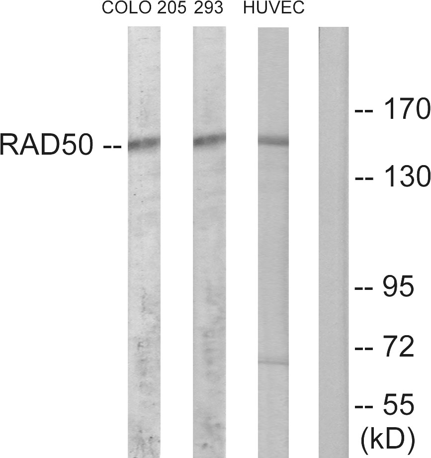
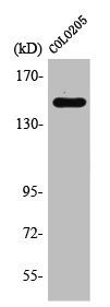
![Various whole cell extracts (30 μg) were separated by 5% SDS-PAGE, and the membrane was blotted with Rad50 antibody [13B3] (GTX70228) diluted at 1:1000. The HRP-conjugated anti-mouse IgG antibody (GTX213111-01) was used to detect the primary antibody.](https://www.genetex.com/upload/website/prouct_img/normal/GTX70228/GTX70228_43388_20181109_WB_23062718_835.webp "Various whole cell extracts (30 μg) were separated by 5% SDS-PAGE, and the membrane was blotted with Rad50 antibody [13B3] (GTX70228) diluted at 1:1000. The HRP-conjugated anti-mouse IgG antibody (GTX213111-01) was used to detect the primary antibody.")


Rad50 antibody [13B3] detects Rad50 protein at nucleus in CAL 27 xenograft by immunohistochemical analysis. Sample: Paraffin-embedded CAL 27 xenograft. Rad50 antibody [13B3] (GTX70228) diluted at 1:200.
Antigen Retrieval: Citrate buffer, pH 6.0, 15 min
Rad50 antibody [13B3]
GTX70228
ApplicationsImmunoFluorescence, ImmunoPrecipitation, Western Blot, ChIP Chromatin ImmunoPrecipitation, ImmunoCytoChemistry, ImmunoHistoChemistry, ImmunoHistoChemistry Paraffin, Other Application
Product group Antibodies
ReactivityHuman, Monkey, Mouse, Rat
TargetRAD50
Overview
- SupplierGeneTex
- Product NameRad50 antibody [13B3]
- Delivery Days Customer9
- Application Supplier NoteWB: 1:500-1:3000. ICC/IF: 1:100-1:1000. IHC-P: 1:100-1:1000. *Optimal dilutions/concentrations should be determined by the researcher.Not tested in other applications.
- ApplicationsImmunoFluorescence, ImmunoPrecipitation, Western Blot, ChIP Chromatin ImmunoPrecipitation, ImmunoCytoChemistry, ImmunoHistoChemistry, ImmunoHistoChemistry Paraffin, Other Application
- CertificationResearch Use Only
- ClonalityMonoclonal
- Clone ID13B3
- Concentration1 mg/ml
- ConjugateUnconjugated
- Gene ID10111
- Target nameRAD50
- Target descriptionRAD50 double strand break repair protein
- Target synonymsNBSLD, RAD502, hRad50, DNA repair protein RAD50, RAD50 homolog, double strand break repair protein
- HostMouse
- IsotypeIgG1
- Protein IDQ92878
- Protein NameDNA repair protein RAD50
- Scientific DescriptionThe protein encoded by this gene is highly similar to Saccharomyces cerevisiae Rad50, a protein involved in DNA double-strand break repair. This protein forms a complex with MRE11 and NBS1. The protein complex binds to DNA and displays numerous enzymatic activities that are required for nonhomologous joining of DNA ends. This protein, cooperating with its partners, is important for DNA double-strand break repair, cell cycle checkpoint activation, telomere maintenance, and meiotic recombination. Knockout studies of the mouse homolog suggest this gene is essential for cell growth and viability. Mutations in this gene are the cause of Nijmegen breakage syndrome-like disorder.[provided by RefSeq, Apr 2010]
- ReactivityHuman, Monkey, Mouse, Rat
- Storage Instruction-20°C or -80°C,2°C to 8°C
- UNSPSC41116161
Datasheet
Related products
Product group Antibodies
Anti-RAD50 AntibodyA97304
ApplicationsWestern Blot, ELISA
ReactivityHuman, Mouse, Rat
- SizePrice
Product group Antibodies
Anti-RAD50 Antibody144-03078
ApplicationsImmunoPrecipitation, Western Blot
ReactivityHuman, Mouse, Rat
TargetRAD50
- SizePrice

Product group Antibodies
Anti-RAD50 Antibody Picoband(r)A00347-2-CARRIER-FREE
ApplicationsFlow Cytometry, ImmunoFluorescence, Western Blot, ELISA, ImmunoCytoChemistry, ImmunoHistoChemistry
ReactivityHuman, Mouse, Rat
TargetRAD50
- SizePrice
Product group Antibodies
RAD50 Recombinant Antibody, AbBy Fluor-488 ConjugatedBSM-61559R-BF488
ApplicationsWestern Blot
ReactivityHuman, Mouse, Rat
TargetRAD50
- SizePrice
Product group Antibodies
RAD50 AntibodyCSB-PA003915
ApplicationsWestern Blot, ELISA, ImmunoHistoChemistry
ReactivityHuman, Mouse, Rat
TargetRAD50
- SizePrice
Product group Antibodies
ApplicationsImmunoPrecipitation, Western Blot, ImmunoCytoChemistry, ImmunoHistoChemistry
ReactivityMouse, Rat
TargetRAD50
- SizePrice
Product group Antibodies

RAD50 AntibodyLS-C402529
ApplicationsWestern Blot, ELISA, ImmunoHistoChemistry
ReactivityHuman, Mouse, Rat
TargetRAD50
- SizePrice

Product group Antibodies
Anti-RAD50 AntibodyHPA052291
ApplicationsImmunoCytoChemistry, ImmunoHistoChemistry
ReactivityHuman
TargetRAD50
- SizePrice
![Non-transfected (–) and transfected (+) HeLa whole cell extracts (30 μg) were separated by 5% SDS-PAGE, and the membrane was blotted with Rad50 antibody [N1N2], N-term (GTX119731) diluted at 1:4000. The HRP-conjugated anti-rabbit IgG antibody (GTX213110-01) was used to detect the primary antibody.](https://www.genetex.com/upload/website/prouct_img/normal/GTX119731/GTX119731_40344_20160901_WB_shRNA_watermark_w_23060519_973.webp)
Product group Antibodies
Rad50 antibody [N1N2], N-termGTX119731
ApplicationsImmunoPrecipitation, Western Blot, ImmunoHistoChemistry, ImmunoHistoChemistry Paraffin
ReactivityHuman, Mouse, Rat
TargetRAD50
- SizePrice
![Mouse tissue extract (50 μg) was separated by 5% SDS-PAGE, and the membrane was blotted with Rad50 antibody [C1C2], Internal (GTX119732) diluted at 1:5000.](https://www.genetex.com/upload/website/prouct_img/normal/GTX119732/GTX119732_40344_20160811_WB_M_testis_w_23060519_827.webp)
Product group Antibodies
Rad50 antibody [C1C2], InternalGTX119732
ApplicationsImmunoPrecipitation, Western Blot, ImmunoHistoChemistry, ImmunoHistoChemistry Paraffin
ReactivityHuman, Mouse, Rat
TargetRAD50
- SizePrice